
Figure 3.
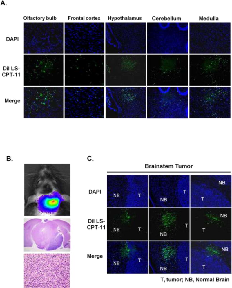
Distribution of fluorescently labeled liposomal CPT-11 by IND in normal brain and brainstem tumor. A. Fluorescent signals were detected from olfactory bulb throughout the different brain regions. B. 1 × 10⁵ luciferase-modified GS2 glioblastoma cells were injected into brainstem in athymic rats using an implantable guide-screw system. Bioluminescence imaging (BLI) shows a corresponding signal from the brainstem tumor (upper). Histologic analysis reveals GS2 tumor growth in the pons (middle: 2 × magnification, lower: 40 × magnification). C. Fluorescent labeled liposomal CPT-11 accumulated in the brainstem 6 hours following IND.