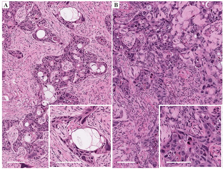
Figure 3.

Histopathologic analyses of excised tumors stained with H&E. A. Moderately differentiated squamous cell carcinoma infiltrating a dense fibroblastic stroma. Numerous pseudocystic structures (white spaces) can be seen characteristic of this squamous cell carcinoma cell line. The inset shows a hyperintense multilocular structure containing cell debris. The proliferating cells are quite abnormal, depicting irregularly shaped and hyperchromatic nuclei. Bars represent 100 μm. B. Moderate to poorly differentiated squamous cell carcinoma infiltrating a hyalinized fibroblastic stroma. Squamous differentiation is focal. The inset shows the atypicity of the proliferating cells and the absence of clear keratinization. Bars represent 100 μm.
