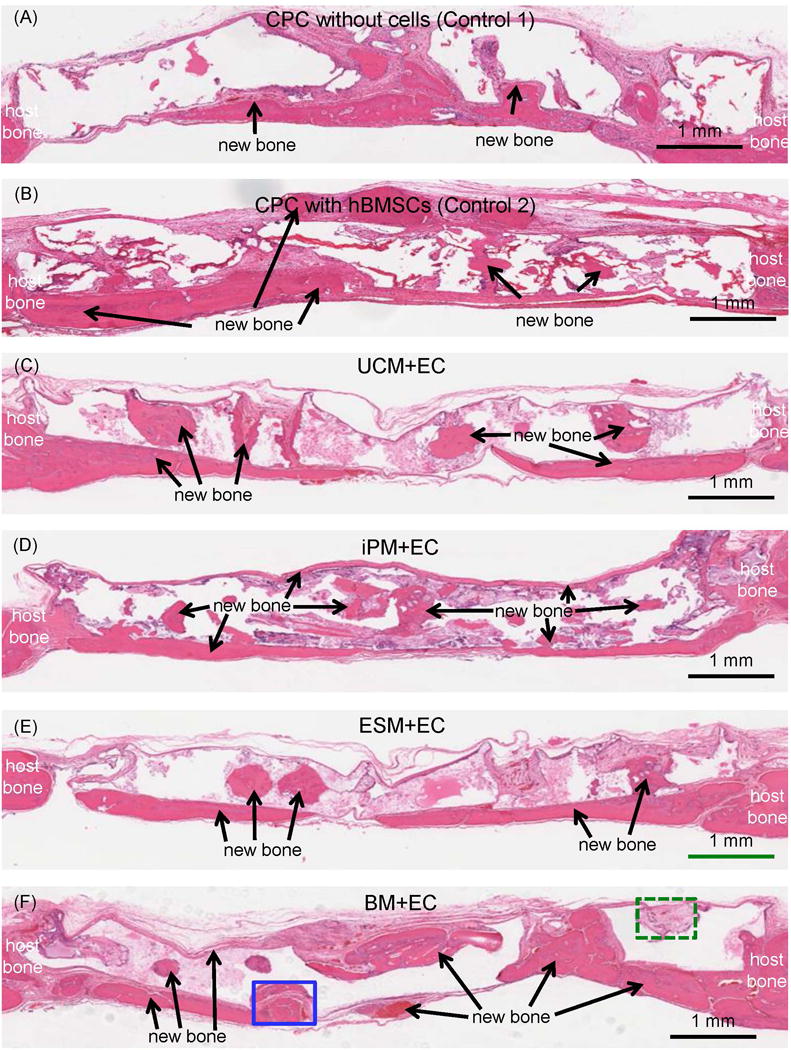
Figure 4.

Typical H&E staining histological images of critical-sized defects in rats at 12 weeks. The group name is listed in the middle of each image. The periosteal side of the defect is on the top, and the dura side is on the bottom, of the image. New bone (arrows) was observed in all groups. The blank areas in the images were CPC which became blank in H&E staining images due to decalcification.
