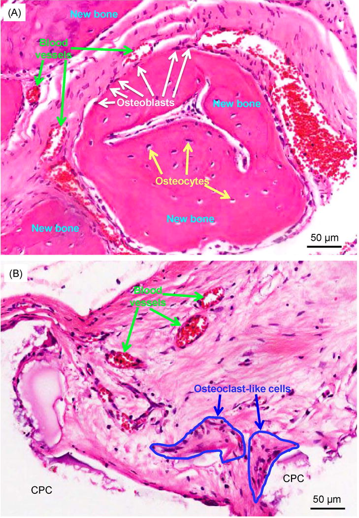
Figure 5.

High magnification images showing typical details in defects: (A) High magnification image of the solid-line rectangle in Fig. 4F, and (B) dotted-line rectangle in Fig. 4F. In (A), osteoblasts with a spindle morphology (white arrows) were found around new bone. Osteocytes (yellow arrows) were found inside new bone. New blood vessels (green arrows) were found around the new bone area. In (B), osteoclast-like multinuclear giant cells (encircled by blue lines) surrounded the CPC surface in the resorption lacunae. The CPC areas were blank in the H&E staining images due to decalcification.
