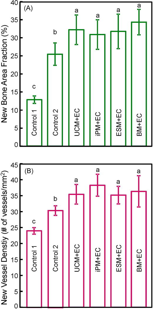
Figure 6.

Quantitative histomorphometry results for critical-sized cranial defects in rats at 12 weeks: (A) Percentage of new bone area, and (B) blood vessel density (mean ± sd; n = 5). Macroporous CPC-RGD containing cocultured cells showed greater new bone area and new blood vessel density than scaffold with hBMSCs alone (control 2) or without cells (control 1). Bars with dissimilar letters indicate significantly different values (p < 0.05).
