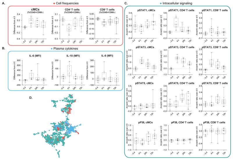
FIGURE 2. Abdominal surgery elicits canonical immune responses.
Representative changes in cell frequency, intracellular signaling, and cytokine plasma concentration are shown. The same changes have previously been described in patients undergoing other types of surgery (1,31). Depicted are changes observed in patients from the control group (n = 11). Changes are calculated as the difference in cell frequency (%CD45+CD66− cells), intracellular signaling activity (Arcsinh transform of mass cytometry signal), and cytokine plasma concentration (mean fluorescence intensity, MFI) between peri-operative time points (−5d, 4h, 24h, 72h) and day 0 (1h before surgery). Box plots represent median and interquartile range. (A) Classical monocytes (cMCs) increased, CD4+ T decreased, and CD8+ T cells decreased in frequency after surgery. (B) Increased signal transducer and activator of transcription (STAT) 1, STAT3, and STAT5 phosphorylation in cMCs, CD4+ T, and CD8+ T cell subsets (4h, 24h, or 72h depending on cell type) and increased phosphorylation of mitogen-activated protein kinase P38 (24h, 72h) in cMCs (but not in CD4+, or CD8+T cells) also recapitulate sentinel cell-type specific signaling changes previously observed in patients undergoing orthopedic surgery (1). (C) Plasma concentrations of IL-6, IL-8, and IL-10 increased after surgery. (D) The entire dataset composed of 316 immune features is represented by a minimum spanning tree emphasizing the correlations between the tree categories of immune features (red dots, cell frequencies, green dots, cell signaling; blue dots, plasma cytokines).

