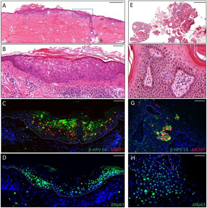
Figure 2.
Immunohistochemical staining for β-HPV E4, MCM7, and ΔNp63 in one AK lesion (A–D) and one seborrheic keratoses (SK) lesions (E–H) derived from a male KTR. (A) H&E staining of a representative tissue section (scale bar: 1,000 μm) from one AK lesion on the back of the patient. (B) Magnification of the region highlighted by the blue box shown in A (scale bar: 100 μm). (C) Immunofluorescence staining for β-HPV E4 (green) and MCM7 (red) of the same tissue section shown in (A,B). (D) Immunofluorescence staining for ΔNp63 (green) of a serial section. The white dotted line indicates the basal layer. (E) H&E staining of a representative tissue section (scale bar: 1,000 μm) from one SK lesion on the forearm of the patient. (F) Magnification of the region highlighted by the blue box shown in E (scale bar: 50 μm). (G) Immunofluorescence staining for β-HPV E4 (green) and MCM7 (red) of the same tissue section shown in (E,F). (H) Immunofluorescence staining for ΔNp63 (green) of a serial section. Sections (C,D,G,H) were counterstained with DAPI (blue) to visualize cell nuclei.

