Abstract
Cocaine is one of the powerful addictive drugs, widely abused in most Western countries. Because of high lipophilic nature, cocaine easily reaches various domains of the central nervous system (CNS) and triggers different levels of cellular toxicity. The aim of this investigation was to reproduce cocaine toxicity in differentiated PC12 cells through quantitative knowledge on biochemical and cytotoxicity markers. We differentiated the cells with 0.1 μg/ml nerve growth factor (NGF) for 5 days, followed by treatment with cocaine for 48 h at in vivo and in vitro concentrations. Results indicated that cocaine at in vivo concentrations neither killed the cells nor altered the morphology, but decreased the mitochondrial membrane potential that paralleled with increased lactate and glutathione (GSH) levels. On the other hand, cocaine at in vitro concentrations damaged the neurites and caused cell death, which corresponded with increased reactive oxygen species (ROS) generation, plasma membrane damage, and GSH depletion with no detectable nitric oxide (NO) level. While direct understanding of cocaine and cell interaction under in vivo animal models is impeded due to high complexity, our present in vitro results assisted in understanding the onset of some key events of neurodegenerative diseases in cocaine treated neuronal cells.
Introduction
Neuronal development, which involves generation, migration, and differentiation of neurons, is essential for a complete and functional nervous system. Similarly, neuronal outgrowth1, branching2, and retraction3 play an important role in neuronal networking process at embryonic and adult stages. Injury to such neurons by external insults can affect neuronal structure and networking system, and at times can cause immediate neuronal death4. Studies showed that certain external insults like substances of drug abuse can induce changes in the structural integrity of neurons and damage their networking processes5–7.
Cocaine is one of the widely abused drugs that cause psycho-stimulatory effects in the central nervous system (CNS). It elevates the mood of addicts initially but leads to severe psychological disorders8, -such as depression9, anger, aggressiveness, and paranoia10 due to imbalance of neurotransmitters. Irrespective of route of intake, cocaine consumption causes severe negative effects in the body such as increased blood pressure and cytotoxicity in all vital organs of the body like heart and kidneys11–13. In the CNS, cocaine was shown to induce death of dopaminergic neurons14. Owing to its lipophilic and hydrophilic nature, cocaine easily crosses placenta15, thus its use by pregnant women could lead to various complications during fetal development16 or induce abortion17, or result in premature labor.
Very few reports are available for the quantification of cocaine effects on neurite outgrowth, morphological changes or neuronal loss under in vitro conditions. In this study, we investigated whether cocaine-induced changes on the structural integrity of neurons and neurites observed in vivo could be reproduced in cell cultures for better understanding and quantification of those changes. In addition, we also evaluated several biochemical changes both at in vivo pharmacological (low) and in vitro concentrations (high) of cocaine. At pharmacological concentrations, dopamine (DA) level, general mitochondrial activity, membrane potential, lactate release, and glutathione (GSH) level were measured (Biochemical markers), while at in vitro concentrations, we measured cytotoxicity markers such as production of reactive oxygen species (ROS), and lactate dehydrogenase (LDH) release, GSH level, and nitric oxide (NO) generation. We employed rat pheochromocytoma PC12 cells as a model culture in this study.
Results
Differentiation
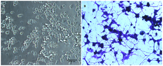
PC12 cells usually grow as floating aggregates in culture medium. In our study, undifferentiated PC12 cells appeared oval to round shape (Fig. 1A). When exposed to NGF at 0.1 μg/ml for five days, the post mitotic cells attached to the collagen coated plates and showed the de-nova neurite outgrowth. Profusely differentiated cells exhibited clear signs of bi or tri polar neurites which appeared slender, mostly straight but branched at some areas with sharp edges (Fig. 1B). In addition, the neurites formed intercellular connections at several areas, demonstrating one of the important features meant for communication by the differentiated neurons. The cell-body mostly appeared as polygonal.
Figure 1.
Morphological features of PC12 cells. Undifferentiated cells grow as round aggregates (A) in the medium (unstained). NGF exposed cells (B) developed extensive growth of neurites (stained with crystal violet dye) with intercellular junctions. Cells were photographed under an inverted phase contrast microscope with 20x objective. Scale bar: 0.06 mm.
Neuronal characteristics
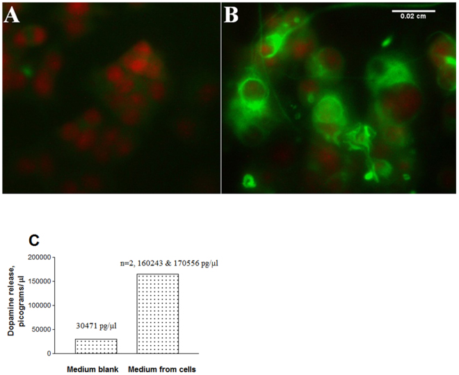
It is well known that differentiated PC12 cells exhibit neuronal phenotype18. To confirm the neuronal characteristics under our experimental conditions, we stained both undifferentiated and differentiated cells for the presence of neurofilaments, which are the intermediate filaments found only in neurons. The red structures in propidium iodide (PI) stained cells represent the nuclei. While lack of staining in undifferentiated cell indicated the absence of neurofilaments (Fig. 2A), the presence of significant staining (green) in NGF-differentiated cells clearly showed the neurofilaments (Fig. 2B).
Figure 2.
Neuronal-like characteristics of differentiated PC12 cells. After fixation and permeabilization, the undifferentiated (A) or NGF exposed (B) cells in 96-well plates were stained with primary rabbit anti-rat NF 200 kD and secondary goat anti-rabbit Alexa Fluor® 488. Cells were counterstained for nuclei with PI and photomicrographed using an inverted fluorescent microscope with a 25x objective. Scale bar: 0.02 mm. For dopamine analysis (C), the medium (50 μl) from differentiated cells was mixed with 100 μl 0.1 N PCA and analyzed by reverse phase HPLC C-18 ODS column at an isocratic flow rate of 1 ml/min.
Differentiated PC12 cells behave as dopaminergic neurons18 and release detectable amount of DA in culture medium. When we analyzed for DA from the medium of differentiated cells by HPLC, it was found that cells released a high level of DA (Fig. 2C). For instance, the amount of DA in the medium blank was 30471 pg/μl, while the medium from differentiated cells contained an average of 165399.5 ± 5156.5 pg/μl DA, -an increase of 5.4 folds. Based on the presence of neurofilaments, neurite outgrowth and DA release, it is clear that the differentiated PC12 cells exhibited neuronal characteristics.
Effect of cocaine at in vivo pharmacological doses on different cellular parameters
Cocaine concentrations in brain cells of drug addicts ranged from nano- to micromolar19. In view of this, we initially treated the differentiated cells with cocaine at various lower concentrations (0.001, 0.01, 0.05, 0.1, 0.5 and 1 mM final) for 48 h to simulate a wide range of in vivo pharmacological doses and evaluated for cell viability. The data clearly indicated that cocaine did not induce significant (n = 4, P = 0.99) cell death at any of these doses compared to the control (Fig. 3A). Similarly, the cell morphology and the extent of neurite branching and connections appeared the same both in the control and at 1 mM cocaine (Fig. 3B). Black arrows point the interneurite connections.
Figure 3.
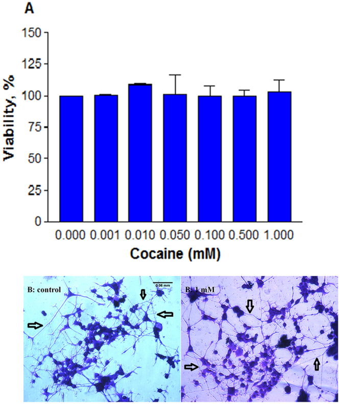
Effect of pharmacological doses of cocaine on viability and morphology. Differentiated cells were treated with PBS (control) or cocaine for 48 h in 96-well plates. Crystal violet stained cells (n = 4, P = 0.99) were assessed for viability (A); morphology of control and 1 mM cocaine treated cells was photographed under an inverted phase contrast 1X-70 Olympus microscope with 20x objective (B), showing neurite connections with black arrows. Data are expressed as mean ± SEM, insignificant compared to the PBS control. Scale bar 0.06 cm.
Since differentiated PC12 cells behaved like dopaminergic neurons, we then studied the effect of cocaine on DA level. The cells were treated with cocaine at 0.075, 0.1, 0.125, 0.25, 0.5 and 1 mM for 48 h. Data obtained by HPLC analysis indicated that the amount of DA at any cocaine treatment was significantly (n = 3, P = 0.03) higher than the control cells (Fig. 4A). There was a dose-dependent increase in DA up to 0.25 mM cocaine, and thereafter a decreasing trend was observed. Compared to the control (100%), the average release in DA was (±SEM) 218.3 ± 8.3, 238 ± 15.1, 234.5 ± 19.1, 221.3 ± 10.2, 226.6 ± 35.9, and 192 ± 55.1% at 0.075, 0.1, 0.125, 0.25, 0.5 and 1 mM cocaine respectively.
Figure 4.
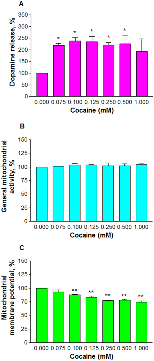
Effect of pharmacological doses of cocaine on cell biochemistry. Differentiated cells were treated with PBS (control) or cocaine for 48 h in 96-well plates. The medium (50 μl) was mixed with 100 μl 0.1 N PCA and analyzed for dopamine release (n = 3, *P = 0.03) by HPLC (A) or the cells were mixed with 10 μl of MTS (n = 3, P = 0.87) 1 h prior to the end-point for general mitochondrial activity (B) or stained with Rh 123 (n = 8, **P = 0.0001) for mitochondrial membrane potential (C). Data are expressed as mean ± SEM, significant compared to the PBS controls.
Next, we evaluated the role of cocaine on the general mitochondrial activity. For this purpose, the cells were treated with cocaine at 0.075, 0.1, 0.125, 0.25, 0.5 and 1 mM for 48 h. The results clearly indicated that cocaine treatment did not interfere (n = 3, P = 0.87) in the general mitochondrial activity of the cells (Fig. 4B). We then measured the mitochondrial membrane potential using the fluorescence probe Rh 123 (10 μM). This lipophilic dye permeates mitochondria easily and interacts with the inner membrane at micro molar concentrations. The accumulation in mitochondria is proportional to membrane potential. The data clearly indicated that cocaine treatment significantly decreased (n = 8, P = 0.0001) the mitochondrial membrane potential in the cells (Fig. 4C). Compared to the control (100%), the average decrease of the membrane potential was (±SEM) 92.9 ± 3.5, 88 ± 0.6, 83.3 ± 1.9, 77.2 ± 1.1, 77.9 ± 1.7, and 74.7 ± 1.9% at 0.075, 0.1, 0.125, 0.25, 0.5 and 1 mM cocaine respectively.
Disruption in mitochondrial potential usually switches the cells to anaerobic glycolysis for survival. Under such condition, pyruvate is oxidized to lactate and released into the medium. In order to study whether loss in membrane potential (Fig. 4C) resulted in the release of lactate, we treated the cells with cocaine at 0.25, 0.5 and 1 mM for 48 h. It was observed that cocaine treatment caused a significant (n = 8, P = 0.001) release of lactate compared to the control cells (Fig. 5A). No significant release of lactate was detected with cocaine treatments at 0.25 mM or lower. Compared to the control (100%), the average release of lactate was (±SEM) 104 ± 3.2, 184.5 ± 20.1 and 201.3 ± 14.9% at 0.25, 0.5 and 1 mM cocaine respectively. Under this situation, we sought to know the role of cocaine on GSH level. The cells were treated with 0.075, 0.1, 0.125, 0.25, 0.5 and 1 mM cocaine for 48 h. There was a dose-dependent significant (n = 3, P = 0.041 to 0.013) increase in GSH level at all treatments compared to the control (Fig. 5B). Compared to the control (100%), the average increase of GSH in the cells was ( ± SEM) 135.8 ± 2.4, 150.5 ± 6.5, 146 ± 9.9, 165.2 ± 4.9, 147.1 ± 12 and 139.9 ± 8.3% at 0.075, 0.1, 0.125, 0.25, 0.5 and 1 mM cocaine respectively.
Figure 5.
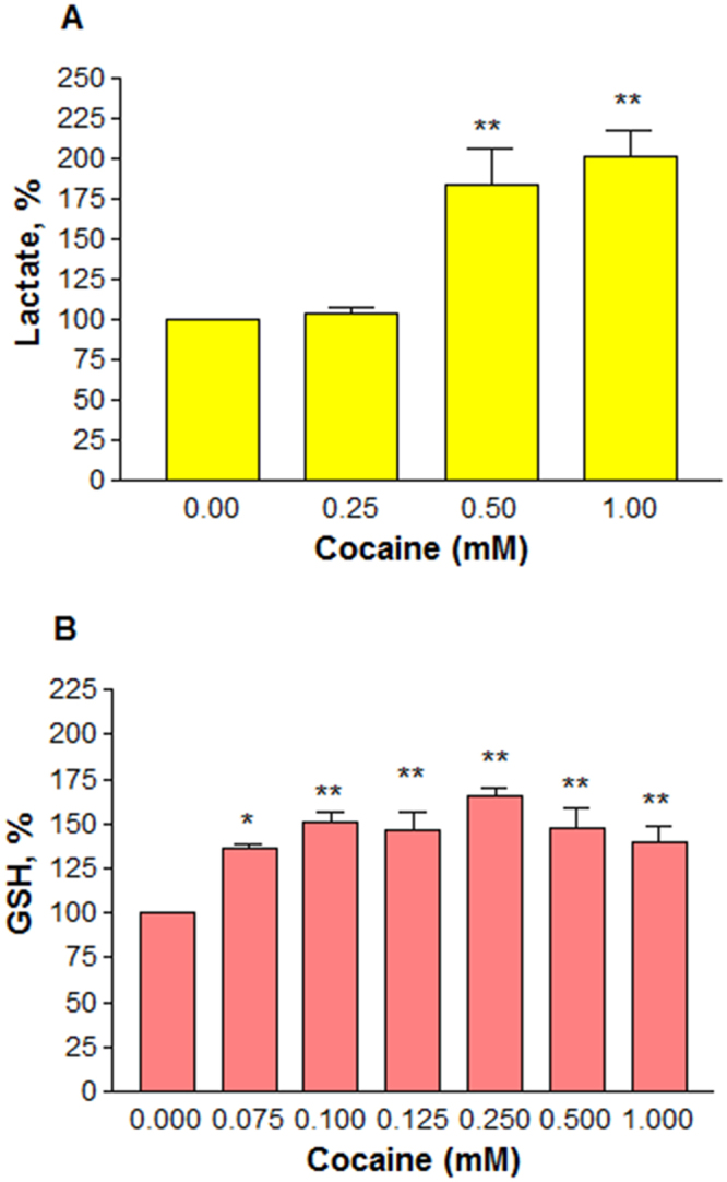
Effect of pharmacological doses of cocaine on the levels of lactate (A) or GSH (B). Differentiated cells in phenol-red free medium were treated with PBS (control) or cocaine for 48 h. Kit reagent (20 μl/well) was added for lactate measurement (n = 8, **P = 0.001), or fixed cells were evaluated for GSH level (n = 3, *P = 0.041 or **P = 0.013). Data are expressed as mean ± SEM, significant compared to the PBS controls.
Effect of cocaine at in vitro doses
Because there was no severe cytotoxic effect of cocaine on cells at different pharmacological concentrations, cocaine doses in the subsequent studies were increased. Based on several in vitro studies20–23, we tested the cells at 2, 3 and 4 mM cocaine for 48 h and evaluated for several cellular parameters as shown below:
Cell viability, ROS and membrane integrity
Higher doses of cocaine caused a significant (n = 12, P = 0.001) dose-dependent decrease in the cell viability (Fig. 6A). For instance, compared to the control (100%), the average decrease in the viability (±SEM) at 2, 3, and 4 mM cocaine was 61.2 ± 1.9, 41.8 ± 2.7, and 22.8 ± 1.8, respectively. The cocaine lethal concentration, LC50, where 50% cells were killed, was found to be 2.605 mM. Neurite elongation was also decreased significantly with increasing cocaine concentrations. For instance, quantification of cellular extensions greater than 2-body diameter in length24 confirmed that cocaine treatment indeed caused a significant (n = 8, P = 0.001) dose-dependant decrease in neurite length (Fig. 6B), shown in red arrows (Fig. 6C). Compared to the control (100%), the average neurite outgrowth (±SEM) at 2, 3 and 4 mM cocaine was 41.3 ± 1.9, 23.8 ± 1.1 and 14.3 ± 1.9%, respectively.
Figure 6.
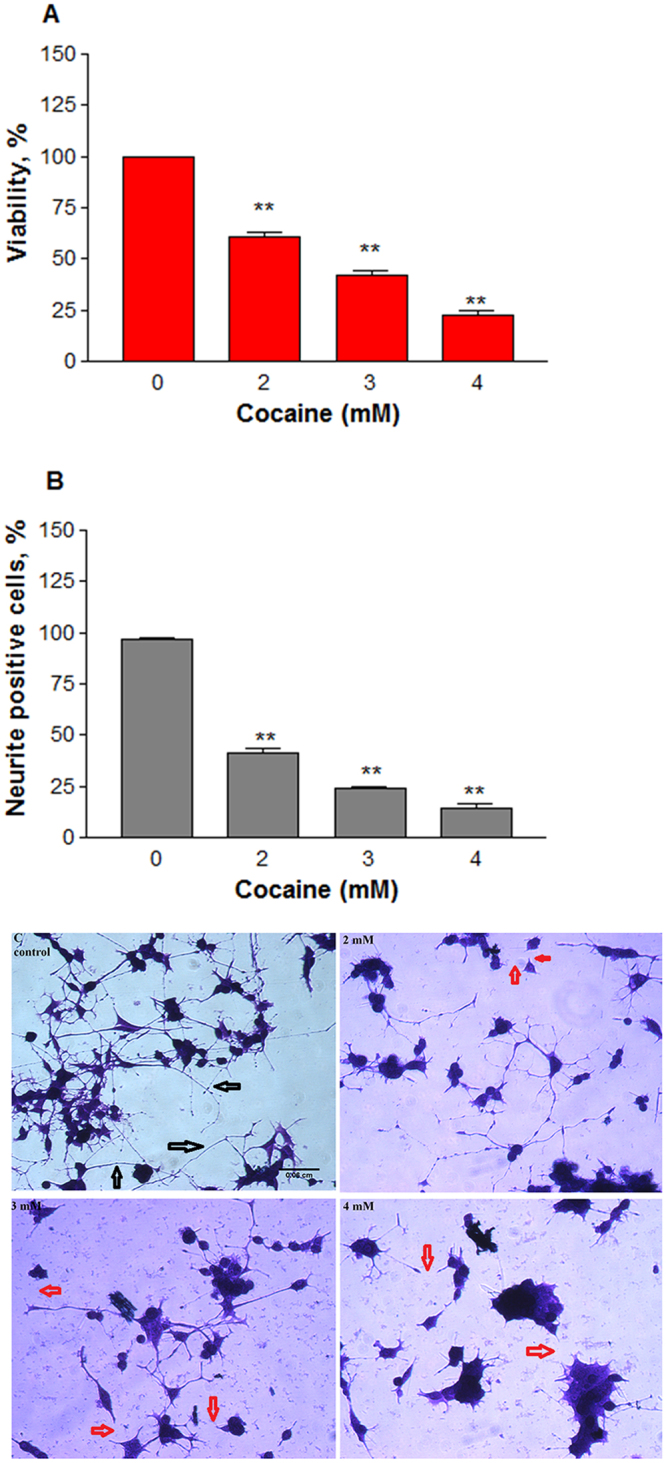
Cytotoxic effect of cocaine at higher doses. Differentiated cells in phenol-red free medium were treated with PBS (control) or cocaine for 48 h. Cell viability (A) was assessed (n = 12, **P = 0.001) by the crystal violet dye uptake method; neuronal extensions greater than 2-body diameter in length of 100 cells per well at four randomly chosen field areas were measured (n = 8, **P = 0.001) and quantified (B); the crystal violet stained cells were photographed under an inverted phase contrast 1X-70 Olympus microscope with 20x objective (C), showing neurite connections with black arrows or disconnected neurites upon cocaine treatments with red arrows. Data are expressed as mean ± SEM, significant compared to the PBS controls. Scale bar 0.06 cm.
We speculated that ROS could be the reason for decrease in the cell viability due to its interaction with mitochondria. So, after treating the cells with cocaine at 2, 3 and 4 mM for 48 h, the ROS level was measured by staining the cells with the fluorescence probe 2′,7′ –dichloro fluorescin diacetate. It was found that there was a significant (n = 12, P = 0.0003) dose-dependent increase in ROS level compared to the control cells (Fig. 7A). The average increase in ROS at 2, 3 and 4 mM cocaine was (±SEM) 135.2 ± 12.1, 148.2 ± 11.3 and 154.2 ± 5.3%, respectively compared to the control (100%). Since high level of ROS can compromise the cell membrane integrity, we then measured the release of cytosolic LDH into the media. A dose-dependent significant (n = 10, P = 0.001) increase in LDH was observed (Fig. 7B). For instance, the average LDH increase at 2, 3, and 4 mM cocaine was (±SEM) 130.7 ± 3.1, 133.4 ± 3.7 and 137.3 ± 8.2%, respectively compared to the control.
Figure 7.
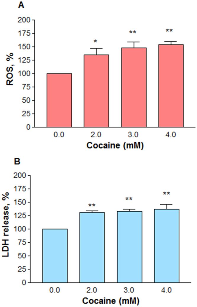
Effect of higher doses of cocaine on ROS generation and membrane integrity. Differentiated cells in phenol-red free medium were treated with PBS (control) or cocaine for 48 h. ROS generation in the cells loaded with DCFDA probe (10 μM) was measured (n = 12, **P = 0.0003) with the excitation and emission filters set at 485 nm and 530 nm respectively in a fluorescence micro plate reader (A); membrane integrity was determined by measuring LDH release (B) from the cells (n = 10, **P = 0.001) at 490 nm in a micro plate reader. Data are expressed as mean ± SEM, significant compared to the PBS controls.
Decreased GSH level
Glutathione is one of the most available antioxidant substances in the cells. In order to explore the potential role of high cocaine concentrations on total glutathione levels, the PC12 cells were treated with different cocaine concentrations (2–4 mM) for 48 h. It was found that cocaine treatment caused a significant (n = 7, P = 0.0004) decrease in GSH level compared to the control (Fig. 8A). The decrease in GSH levels was (±SEM) 25 ± 7.1, 34.1 ± 5.4 and 33.9 ± 5.8% of the control value (100%) at 2, 3 and 4 mM cocaine, respectively.
Figure 8.
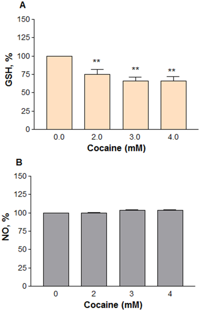
Effect of higher doses of cocaine on GSH or NO level. Differentiated cells in phenol-red free medium were treated with PBS (control) or cocaine for 48 h. GSH (A) level in fixed cells (n = 7, **P = 0.0004) was measured as per established procedure at 412 nm in a micro plate reader; for NO detection (B), 100 μl of medium was transferred into a new 96-well plate (n = 9, P = 0.6) and mixed with equal volume of Griess reagent, followed by 10 min incubation in the dark. The absorbance readings at 540 nm were measured in a micro plate reader. Data are expressed as mean ± SEM and compared to the PBS controls.
Lack of inflammatory response
Overproduction of nitric oxide indicates one of the inflammatory responses. While nitric oxide (NO) is unstable at physiological pH, its interaction with air eventually leads in the formation of a more stable nitrite that can be detected easily by Greiss reagent. In order to understand the role of cocaine on inflammation, we treated the cells with cocaine at 2, 3 and 4 mM doses for 48 h. The cells were not challenged with bacterial lipopolysaccharide or γ interferon during the treatment period because differentiated PC12 cells have nNOS25,26, and we wanted to know if cocaine induces this enzyme for excessive NO production. The data indicated clearly that in comparison to the control, cocaine at any concentration did not stimulate or inhibit NO production significantly in the cells (n = 9, P = 0.6,) after 48 h (Fig. 8B).
Discussion
Neurons are the basic components of CNS regarding signal transmission needed for cell-cell communication through dendrites and axons. Under in vitro condition, PC12 cells were routinely employed as a neuronal model to study neurites27–29. These cells were also used extensively as a model to study cocaine effects30–32. Originally, this cell line was obtained from the rat adrenal pheochromocytoma33, and maintained as undifferentiated aggregates. Upon differentiation with NGF, the cells exhibit phenotypes of both acetylcholine and dopaminergic neurons18. These cells were also shown to express dopamine transporters (DAT)32. Under our standard experimental conditions, NGF-stimulated PC12 cells showed extensive differentiation on collagen coated plates (Fig. 1B) compared to the un-stimulated cells (Fig. 1A). We confirmed the neuronal phenotype of these cells by staining for neurofilaments (Fig. 2B) compared to un-stimulated cells (Fig. 2A). Furthermore, we found that the differentiated cells released high level of DA (Fig. 2C) compared to the medium blank. Based on these observations (Fig. 2B, and C), our results confirmed earlier report18 that differentiated PC12 cells behaved like dopaminergic neurons of mammalian CNS.
Cocaine, a widely abused drug, not only shows psycho-stimulatory effects, but also causes severe damage to various brain cells through several toxic responses. Cocaine concentrations to elicit pharmacological response in human addicts have ranged from nano to micromolar19 but under in vitro cell culture studies, they extended into milli molar range20–22,34. In order to study cocaine effects on structural changes in cells, we employed the concentrations spanning from nano, micro and milli molar range. While the selection of lower concentrations in our study was based on earlier reports19, the selection of in vitro (higher) concentrations of cocaine was based on the LC50 and EC50 values found in the literature21,22,35,36 and on our own assessments of the minimal concentrations of cocaine needed to induce detectable changes under in vitro studies23,37,38. The use of high concentrations such as 8.8 mM39 or 10 mM20,21 or even 13 mM40 cocaine under in vitro conditions is not atypical. Compared to these high doses, we believe that cocaine concentrations (2–4 mM) in our study were within the realm of experimental research. When tested at pharmacological concentrations, cocaine did not induce significant cell death (Fig. 3A) nor did it damage the neurites (Fig. 3B).
In the CNS, cocaine is known to prevent the reuptake of DA by blocking DA transporters on pre-synaptic neurons41. Since this inhibition doesn’t prevent further release of DA from pre-synaptic neurons, its concentration at the synaptic cleft could increase several fold42,43. In our study, a dose-dependent increase in DA (Fig. 4A) suggests that cocaine caused an inhibition of DA reuptake, resulting its accumulation in the medium. Previous studies showed that cocaine action in PC12 cells was through DAT32 which was further confirmed by a dose-dependent inhibition of dopamine reuptake with cocaine treatment in PC12 cells44. Although cocaine treatment up to 1 mM did not change the general mitochondrial activity (Fig. 4B) in cells, it significantly affected the membrane potential (Fig. 4C), an observation consistent with previous reports on the loss of membrane potential with cocaine treatment in cells21,45 or C6 astroglia-like cells46 or in cells outside of CNS47. These observations suggest that mitochondria are the primary targets of cocaine-induced toxicity in various cell-types. While a stable membrane potential is essential for normal functioning of mitochondria, its loss due to cocaine exposure causes the cells to switch to anaerobic respiration where pyruvate is converted to lactate. A dose-dependent increase in lactate production in our study (Fig. 5A) confirms anaerobic conditions in cocaine treated cells. We did not observe any ROS in cells treated up to 1 mM cocaine (data not shown). The reason for this is attributed to high GSH level (Fig. 5B) or other antioxidants48,49 in cocaine treated cells. Non detection of ROS and lack of cell death (Fig. 3A) up to 1 mM cocaine imply that the increased GSH level (Fig. 5B) through increased glutathione peroxidase48 was sufficiently enough to counteract oxidative damage. Lack of both cell death (Fig. 3A) and damage to neurite outgrowth (Fig. 3B) with cocaine at in vivo concentrations in our study suggests that one-time cocaine consumption by humans that yields the pharmacological (lower) doses in the brain may not affect the neuronal viability and outgrowth. However, since addicts consume cocaine several times, it is possible that neuronal viability and outgrowth are affected in the long-run. For instance, post-mortem examination of several long-term cocaine addicts showed loss of dopaminergic neurons in the striatal and mid-brain14. Some of the factors associated with neuronal loss could be elevated ROS and lipid peroxidation50.
Cocaine did not cause cell death up to 1 mM (Fig. 3A). In order to observe specific changes in the cells, it was necessary to increase the treatment concentrations of cocaine which are within acceptable range for other in vitro studies20–22,34. Studies conducted in non-neuronal cells such as C6 astroglia-like cells indicated that treatment with cocaine at higher concentrations caused drastic cell death23,46. Consistent with these observations, when differentiated PC12 cells were treated with similar cocaine concentrations, we observed a significant decrease in the viability (Fig. 6A). These results corroborated with earlier reports, where cocaine cytotoxicity in PC12 cells was confirmed by different assays51,52; however, the extent of cocaine cytotoxicity was not quantified in those reports. In our study, the LC50 of cocaine was found to be 2.605 mM at 48 h end point. This value was significantly lower than the LC50 in C6 astroglia-like cells for cocaine at a 48 h endpoint study46 (3.794 mM). The lower LC50 value in PC12 cells may suggest that these cells were more sensitive to cocaine compared to C6 astroglia-like cells.
Cocaine treatment at higher concentrations significantly decreased the neurite length (Fig. 6B) and resulted in the loss of intercellular connections. Similar observations were reported earlier in rat53 and neuronal cultures treated with cocaine54. It is well known that treatment of cells with drug abuse substances often results in cytoplasmic vacuolation, an indication of cell injury. For instance, our previous studies with C6 astroglia-like cells indicated that cocaine exposure caused extensive vacuolation23,46, which were in agreement with reports on different cell-types55–58 Interestingly, the phenomena of vacuolation was not observed in differentiated PC12 cells when treated with cocaine in our study.
Compounds that interfere with mitochondrial function usually cause oxidative stress in cells23. Since cocaine interacts with mitochondria46 and impairs the energy metabolism, its toxicity in cells is often associated through oxidative stress. A dose-dependent increase in ROS level (Fig. 7A) in treated cells not only confirmed cocaine’s interaction with mitochondria but also indicated its role in the loss of cell membrane integrity (Fig. 7B). Cocaine-induced ROS generation was reported earlier in different cell-types or animals23,48,59,60. ROS is also generated from rapid auto oxidation of DA released from differentiated PC12 cells61,62; similarly, dysfunctional mitochondria also generate ROS in cells63. However, since DA in our study was released into culture medium, the extracellular auto oxidation of DA is not the source of ROS generation because it cannot be detected by our ROS method. Therefore, the observed ROS (Fig. 7A) with cocaine treatment at higher concentrations ought to be intracellular only. As mitochondria are the main source of ROS formation64,65, the intracellular ROS generation (Fig. 7A) suggests its mitochondrial origin. The release of lactate (Fig. 5A) and loss in membrane potential (Fig. 4C) in our study further supports the dysfunctional state of mitochondria for ROS generation. Similar results of ROS generation were reported in undifferentiated PC12 cells treated with cocaine66. Since ROS was the cause of cell death in our study, it is speculated that mitochondria were one of the pharmacological targets recruited at higher cocaine concentrations. Increased ROS with cocaine treatment paralleled with the decrease in GSH amount in cells (Fig. 8A), suggesting that ROS was the main cause of cell death (Fig. 6A). This observation was consistent with previous reports where cocaine treatment increased the oxidative damage through decreased antioxidants in the cells59. The increase (Fig. 5B) and decrease (Fig. 8A) of GSH level with low and high concentrations of cocaine respectively show the biphasic effect of cocaine in the cells.
Damaged or dysfunctional neurons could trigger inflammation by other non-neuronal cell types -like microglia and astrocytes in the CNS. So, we next investigated whether damage or death of differentiated PC12 cells due to cocaine treatment triggered the release of NO, one of the indicators of inflammatory response. It was found that there was no significant change in NO level between the control and cocaine treated groups under our experimental conditions (Fig. 8B). Lack of induction of NO in our study with cocaine was consistent with earlier reports on different cell types67. Since excessive generation of NO is generally associated with cell cytotoxicity, lack of high NO release with cocaine treatment in our study indicates that NO was not the cause of cocaine-induced cytotoxicity.
Conclusion
Dysfunctional mitochondria68, or changes in neuronal structure69,70 or loss in neurite-connections could lead to psychiatric illnesses -such as depression9, anger, aggressiveness, and paranoia10. The findings of dysfunctional mitochondria (Fig. 4C) and neurite-damage (Fig. 6B,C, 2–4 mM) in our study appear to suggest that these changes could be some of the contributing factors for psychiatric illnesses and altered psychology in cocaine addicts. On the other hand, since unusual glucose oxidation due to dysfunctional mitochondria is a common feature in schizophrenia and Alzheimer’s disease71, the dose-dependent release of lactate (Fig. 5A), a product of glucose oxidation due to dysfunctional mitochondria (Fig. 4C) in our study, points out the possibility that cocaine users may be increasingly at risk for these neurodegenerative disorders. Further studies are required to ascertain this claim.
Prenatal cocaine exposure could impede dendrite-axon connections in fetus and greatly impact the developing brain6. Based on our results (Figs 4C, 6A,B), it can be speculated that the developing fetus could be at risk for neurite damage and damage to neuronal structures if the mother is using cocaine during pregnancy. This may severely affect the brain of developing fetus6. As per estimations, around 0.75 million pregnant women are exposed to cocaine every year72. Compounds which could prevent cocaine-induced neurite damage could play a vital role in reducing the cocaine toxicity to neurons.
Materials and Methods
Materials
RPMI 1640, fetal bovine serum (FBS), penicillin/streptomycin, amphotericin B, phosphate-buffered saline (PBS), Hank’s balanced salt solution (HBSS), and L-glutamine were purchased from Media Tech (Herndon, VA). Horse serum (HS) was obtained from Invitrogen (Waltham, MA). Cocaine-HCl (Ecgonine methyl ester benzoate, MW: 339.8), crystal violet, 2′,7′-dichlorofluorescin diacetate, L-glutaraldehyde, trypan blue, N-1-napthylethylenediamine dihydrochloride (NED), rhodamine (Rh)123 and EDTA (ethylene diamine tetraacetic acid) were supplied by Sigma Chemical Company (St. Louis, MO). All other routine chemicals were of analytical grade.
Cell culture maintenance
Rat pheochromocytoma PC12 cell line was obtained from American Type Culture Collection (Rockville, MD) and maintained as a suspension culture in RPMI 1640 medium supplemented with 2 mM L-glutamine, 5% (v/v) FBS, 10% (v/v) HS, 100 U/ml penicillin, 100 μg/ml streptomycin and 0.25 μg/ml amphotericin B. Cells were grown in a humidified atmosphere containing 5% CO2 in air at 37 °C in an incubator. For sub-culturing, loosely attached cells in the flask were collected and centrifuged at 304 g for 4 min at room temperature. The cell pellet was washed with HBSS under sterile conditions, re-suspended in 1 ml complete medium and passed through 25 gauge needle 4–5 times to obtain singlet cells. An aliquot of it was used for cell counting with 0.4% trypan blue on hemocytometer under a light microscope. Dye stained cells (blue) were counted as dead cells, while dye excluded cells were counted as viable. The actual cell numbers were determined by multiplying diluted times compared with initial cell numbers.
Differentiation of cells
Lyophilized mouse submaxillary gland derived nerve growth factor (NGF, 97% pure, Chemicon International, Inc., Temecula, CA), was reconstituted with sterile DMEM at 0.1 mg/ml, aliquoted and stored at −70 °C. After cell count, cells were diluted in the complete RPMI 1640 medium containing 2.3% FBS, 2.3% HS, 100 ng/ml NGF, and seeded at a low density (2 × 10⁴ cells/0.3 ml per well) in collagen coated 24-well or 96-well sterile culture plates (BD Labware, Bedford, MA). The cells were incubated for five days with the change of NGF containing complete medium once in every 48 h.
Morphology
For gross cellular morphological evaluation, the crystal violet stained cells were observed under an inverted phase contrast IX-70 Olympus microscope (Olympus, Ontario, NY) with 20x objective. Photomicrographs were taken by an ocular video-camera system (MD35 Electronic eyepiece, Zhejiang JinCheng Scientific & Technology Co., Ltd, HangZhou, China) using C-Imaging System Software (Compix Inc. Cranberry Township, PA).
Immunocytochemistry and fluorescence Microscopy
Cells were fixed in 4% paraformaldehyde for 15 min, and subsequently permeabilized in 0.5% triton X -100 prepared in PBS for 15 min. Neurofilament 200 kD was determined using immunocytochemistry in fixed permeabilized cells, after blocking with primary rabbit anti-rat NF 200 kD, conjugated to goat anti-rabbit Alexa Fluor® 488 - and nuclear counterstained with PI. Samples were analyzed photographically using a fluorescent/inverted microscope, CCD camera and data acquisition using ToupTek View (ToupTek Photonics Co, Zhejiang, P.R. China) with a 25x objective.
Dopamine assay
The media (50 μl) from 5 d post seeded or 48 h of cocaine treated cells (0.075, 0.1, 0.125, 0.25, 0.5 and 1 mM) in collagen coated 96-well plates were transferred into new sterile 96-well plates, mixed with 100 μl 0.1 N perchloric acid (PCA) per well and stored at −70 °C until HPLC analysis were performed. On the day of study, the plates were thawed and vortexed gently. From each well, 100 μl of sample was transferred into HPLC vial that contained 700 μl of mobile phase [75 mM sodium phosphate (monobasic), 1.7 mM 1-octanesulfonic acid, 25 μM Na2EDTA, 10% acetonitrile and 0.01% triethylamine, pH adjusted to 3.0 by phosphoric acid]. Dopamine was used as a standard reference (0.5 μg/ml). Dopamine level in the samples was detected by reverse phase HPLC C-18 ODS column (4 mm I.D. 7.6 cm, 3 μm particle size) at an isocratic flow rate of 1 ml/min. The HPLC was equipped with an ESA Model 5200 Coulochem III high-sensitivity analytical electrochemical detector. HPLC analysis was carried out using EZChrom software package by Scientific Software (Pleasanton, CA).
Cocaine treatments
A known amount of cocaine was dissolved in PBS as 1 M stock just prior to the assays. In order to test cocaine toxicity at lower concentrations (0.001, 0.01, 0.05, 0.1, 0.5 and 1 mM final), several working stocks (0.04, 0.4, 2, 4, 20 and 40 mM) were prepared in PBS; similarly, for studies at higher cocaine concentrations (2, 3 and 4 mM final), different working stocks (80, 120 and160 mM) were prepared in PBS. Cocaine samples were always added in a minimum volume of 5 μl per well to prevent pH alterations in culture medium46. Final volume in each well was 200 μl. Cells with medium alone or equal volume of vehicle (PBS) in medium served as controls. Cocaine treatments were continued for 48 h at 37 °C, 5% CO2 in the incubator without further renewal of medium with the plates capped in normal fashion.
Evaluation of cell viability
Cytotoxicity of cocaine was evaluated by the dye uptake assay using crystal violet as described previously73. Absorbance was measured at 540 nm in a microtiter culture plate reader (Bio-Tek Instruments Inc, Wincoski, VT). The average absorbance values of controls were taken as 100% cell viability. From the treated and control absorbance values, percent cells killed were determined by the following equation: [1− (T/C)] × 100, where T is average absorbance values of treated cells, and C is average absorbance values of control cells.
Assessment of neurite outgrowth
At the end of 48 h incubation, 400 μl of 0.25% glutaraldehyde was added per well and incubated for 30 min to fix the cells to culture plates. The plates were gently washed three times, and air dried. Then, the cells were stained with crystal violet as described previously74. The cells were observed under an inverted phase contrast IX-70 Olympus microscope with 20x objective. Neuronal length, defined as the cellular extensions greater than 2-body diameter in length24, was measured at four randomly chosen field areas of total 100 cells per well.
General mitochondrial metabolic activity
It was performed as per earlier reports75,76. The cells in complete media without phenol-red were treated with cocaine at 0.075, 0.1, 0.125, 0.25, 0.5 and 1 mM for 48 h in collagen coated 96-well microtiter plates. One hour prior to the end point, 10 μl of 3-(4,5-dimethylthiazol-2-yl)-5(3-carboxymethonyphenol)-2-(4-sulfophenyl)-2H-tetrazolium (MTS, Promega, Madison, WI) was added per well. Absorbance was taken in a micro plate reader at 490 nm.
Mitochondrial membrane potential
Cells in collagen coated 96-well plates were treated with cocaine at 0.075, 0.1, 0.125, 0.25, 0.5 and 1 mM for 48 h. At the end of incubation, cells were stained with laser dye fluorescent probe rhodamine (Rh) 123 (10 μM) for 30 min at 37 °C. After discarding excess dye, the cells were washed, air dried and 100 μl PBS was added per well. The plates were read with the excitation filter set at 485 nm and the emission filter at 530 nm in an automatic reader.
Lactate assay
In order to minimize the serum interaction with lactate reagent (Trinity Biotech, Jamestown, NY), the differentiated cells in collagen coated 96-well plates were treated with cocaine at different concentrations (0.25, 0.5, 1 mM) in reduced serum (0.1% FBS) for 48 h. Lactate was determined by calorimetric method. The content in lactate reagent was dissolved in chromogenic solution consisting of 5.3 mM vanillic acid, 2.9 mM 4-amino antipyrine, and about 4 units of horseradish peroxidase. At the end of treatment period, lactate reagent (20 μl per 200 μl/well) was added, and the plates were incubated at 37 °C until the color was developed (5–10 min). Absorbance was measured at 490 nm in a microtiter culture plate reader.
GSH level
After treating the differentiated cells with cocaine at lower concentrations (0.075, 0.1, 0.125, 0.25, 0.5, 1 mM) or higher concentrations (2, 3 and 4 mM) for 48 h in collagen coated 96-well microtiter plates, the cells were fixed with 0.25% glutaraldehyde for 30 min, followed by three gentle washings and air drying. Total cellular GSH was assayed as per earlier study77. The absorbance was measured at 412 nm in a micro plate reader.
Intracellular reactive oxygen species (ROS)
Cells were seeded on collagen coated 96-well plates in phenol red free medium containing 0.5% each of FBS and HS. Following incubation of cells for 5 d at 37 °C, 5% CO2 in the incubator to facilitate differentiation, cells were treated with cocaine at 2, 3 and 4 mM for 48 h. For measuring the ROS, the cells were stained with 2′,7′ –dichlorofluorescin diacetate (DCFDA, 10 μM final, Sigma Company, St. Louis, MO) for 30 min. After gentle washing and air drying the cells, PBS (100 μl/well) was added. The plates were read with the excitation filter set at 485 nm and the emission filter at 530 nm in an automatic reader (Synergy HTX multimode micro plate reader, BioTek Instruments, Winooski, VT).
Membrane integrity assay
Cell membrane integrity was determined by measuring LDH release from cells with CytoTox 96 non-radioactive assay kit (Promega) as per kit instructions provided by the manufacturer. In brief, the cells in collagen coated 96-well plates containing 0.5% each of FBS and HS in phenol red free medium were treated with various concentrations of cocaine (2, 3 and 4 mM) for 48 h. Then the media (50 μl) was transferred into new 96-well plates and mixed with equal volume of assay substrate. After 30 min incubation, the color intensity was measured using an automatic micro plate reader at 490 nm.
Nitric oxide assay
Nitric oxide production with cocaine treatment (2, 3 and 4 mM) in collagen coated 24-well plates was assessed according to the method reported previously78 in the complete medium lacking phenol red. In brief, at the end of 48 h treatment with cocaine, 100 µl of medium was transferred into a new 96-titer plate and mixed with an equal volume of Griess reagent (1% sulfanilamide/0.1% NED in 5% phosphoric acid) followed by 10 min incubation in the dark. The absorbance readings at 540 nm were measured in a microtiter culture plate reader.
Statistical analysis
All results were presented as mean ± standard error mean (SEM). The data were analyzed for significance by one-way ANOVA and then compared by Dunnett’s multiple comparison tests using GraphPad Prism Software, version 3.00 (GraphPad Software, Inc., San Diego, CA). The test value of P < 0.05 and 0.01 were considered significant and highly significant, respectively. The LC50 value representing the milli molar concentration of cocaine needed to show 50% response was determined from the graphs as per the method described earlier79.
Data availability
The research data obtained and analyzed in this study are available from the corresponding author on request.
Acknowledgements
The authors thank Cheryl A. Fitch-Pye for reading the manuscript and for suggestions. This study was supported by the National Institute on Minority Health and Health Disparities of the National Institutes of Health under award number G12MD007582, P20 MD006738 and the National Institute of General Medical Sciences of the National Institutes of Health under Award Number R25GM107777. The content is solely the responsibility of the authors and does not necessarily represent the official views of the National Institute of Health.
Author Contributions
R.B.B., C.B.G. and S.C.G. conceived and designed research. R.B.B., C.S.B. and E.M. performed research. R.B.B., C.B.G., and E.M. analyzed the data. R.B.B. wrote the paper. All authors gave the final approval and agreed to be accountable for all aspects of the work.
Competing Interests
The authors declare no competing interests.
Footnotes
Publisher's note: Springer Nature remains neutral with regard to jurisdictional claims in published maps and institutional affiliations.
References
- 1.Gomez TM, Spitzer NC. In vivo regulation of axon extension and pathfinding by growth-cone calcium transients. Nature. 1999;397:350–355. doi: 10.1038/16927. [DOI] [PubMed] [Google Scholar]
- 2.Cuntz H, et al. One rule to grow them all: A general theory of neuronal branching and its practical application. PLoS Comput. Biol. 2010;6(8):e1000877. doi: 10.1371/journal.pcbi.1000877. [DOI] [PMC free article] [PubMed] [Google Scholar]
- 3.Luo L, O’Leary DD. Axon retraction and degeneration in development and disease. Annu. Rev. Neurosci. 2005;28:127–156. doi: 10.1146/annurev.neuro.28.061604.135632. [DOI] [PubMed] [Google Scholar]
- 4.Neumann H, et al. Tumor necrosis factor inhibits neurite outgrowth and branching of hippocampal neurons by a rho-dependent mechanism. J. Neurosci. 2002;22:854–862. doi: 10.1523/JNEUROSCI.22-03-00854.2002. [DOI] [PMC free article] [PubMed] [Google Scholar]
- 5.Robinson TE, Kolb B. Persistent structural modifications in nucleus accumbens and prefrontal cortex neurons produced by previous experience with amphetamine. J. Neurosci. 1997;17:8491–8497. doi: 10.1523/JNEUROSCI.17-21-08491.1997. [DOI] [PMC free article] [PubMed] [Google Scholar]
- 6.Robinson TE, Kolb B. Alterations in the morphology of dendrites and dendritic spines in the nucleus accumbens and prefrontal cortex following repeated treatment with amphetamine or cocaine. Eur. J. Neurosci. 1999;11:1598–1604. doi: 10.1046/j.1460-9568.1999.00576.x. [DOI] [PubMed] [Google Scholar]
- 7.Robinson TE, et al. Cocaine self-administration alters the morphology of dendrites and dendritic spines in the nucleus accumbens and neocortex. Synapse. 2001;39:257–266. doi: 10.1002/1098-2396(20010301)39:3<257::AID-SYN1007>3.0.CO;2-1. [DOI] [PubMed] [Google Scholar]
- 8.Haasen C, et al. Relationship between cocaine use and mental health problems in a sample of European cocaine powder or crack users. World Psychiatry. 2005;4(3):173–176. [PMC free article] [PubMed] [Google Scholar]
- 9.Rounsaville BJ, et al. Heterogenicity of psychiatric diagnosis in treated opiate addicts. Arch. Gen. Psychiatry. 1982;39:161–166. doi: 10.1001/archpsyc.1982.04290020027006. [DOI] [PubMed] [Google Scholar]
- 10.Satel SL, Southwick SM, Gawin FH. Clinical features of cocaine-induced paranoia. Am. J. Psychiatry. 1991;148(4):495–498. doi: 10.1176/ajp.148.4.495. [DOI] [PubMed] [Google Scholar]
- 11.Cregler LL, Mark H. Medical complications of cocaine abuse. N. Engl. J. Med. 1986;315(23):1495–1500. doi: 10.1056/NEJM198612043152327. [DOI] [PubMed] [Google Scholar]
- 12.Kanel GC, et al. Cocaine-induced liver cell injury: comparison of morphological features in man and in experimental models. Hepatology. 1990;11:646–651. doi: 10.1002/hep.1840110418. [DOI] [PubMed] [Google Scholar]
- 13.Zimmerman JL. Cocaine intoxication. Crit. Care Clin. 2012;28:517–526. doi: 10.1016/j.ccc.2012.07.003. [DOI] [PubMed] [Google Scholar]
- 14.Little KY, et al. Decreased brain dopamine cell numbers in human cocaine users. Psychiatry Res. 2009;168:173–180. doi: 10.1016/j.psychres.2008.10.034. [DOI] [PubMed] [Google Scholar]
- 15.Ganapathy V, Leibach FH. Human placenta: a direct target for cocaine action. Placenta. 1994;15:785–795. doi: 10.1016/S0143-4004(05)80181-X. [DOI] [PubMed] [Google Scholar]
- 16.Schenker S, et al. The transfer of cocaine and its metabolites across the term human placenta. Clin. Pharmacol. Ther. 1993;53:329–339. doi: 10.1038/clpt.1993.29. [DOI] [PubMed] [Google Scholar]
- 17.Goldstein RA, DesLauriers C, Burda AM. Cocaine: History, social implications, and toxicity-A review. Disease-a-Month. 2009;55:6–38. doi: 10.1016/j.disamonth.2008.10.002. [DOI] [PubMed] [Google Scholar]
- 18.Teng, K. K. & Greene, L. A. Cultured PC12 cells: a model for neuronal function and differentiation in Cell Biology: A Laboratory Handbook (ed J. E Celis) 218–224 (Academic Press, 1994).
- 19.Zheng, F., & Zhan, C. G. Modeling of pharmacokinetics of cocaine in human reveals the feasibility for development of enzyme therapies for drugs of abuse. PloS Comput. Biol. 8, e1002610 PMID: 22844238, 10.1371/journal.pcbi.1002610 (2012). [DOI] [PMC free article] [PubMed]
- 20.Repetto G, et al. Morphological, biochemical and molecular effects of cocaine on mouse neuroblastoma cells culture in vitro. Toxicol. InVitro. 1997;11:519–525. doi: 10.1016/S0887-2333(97)00066-0. [DOI] [PubMed] [Google Scholar]
- 21.Cunha-Oliveira T, et al. Mitochondrial dysfunction and caspase activation in rat cortical neurons treated with cocaine or amphetamine. Brain Res. 2006;1089:44–54. doi: 10.1016/j.brainres.2006.03.061. [DOI] [PubMed] [Google Scholar]
- 22.Cunha-Oliveira T, et al. Differential cytotoxic responses of PC12 cells chronically exposed to psychostimulants or to hydrogen peroxide. Toxicology. 2006;217:54–62. doi: 10.1016/j.tox.2005.08.022. [DOI] [PubMed] [Google Scholar]
- 23.Badisa, R. B. et al. N-acetyl cysteine mitigates the acute effects of cocaine-induced toxicity in astroglia-like cells. Plos One, 10.1371/journal.pone.0114285 (2015). [DOI] [PMC free article] [PubMed]
- 24.Keilbaugh SA, Prusoff WH, Simpson MV. The PC12 cell as a model for studies of the mechanism of induction of peripheral neuropathy by anti-hiv-1 dideoxynucleoside analogs. Biochem. Pharmacol. 1991;42:R5–8. doi: 10.1016/0006-2952(91)90672-R. [DOI] [PubMed] [Google Scholar]
- 25.Peunova N, Enikolopov G. Nitric oxide triggers a switch to growth arrest during differentiation of neuronal cells. Nature. 1995;375:68–73. doi: 10.1038/375068a0. [DOI] [PubMed] [Google Scholar]
- 26.Kawasaki H, et al. Nitration of tryptophan in ribosomal proteins is a novel post- translational modification of differentiated and naïve PC12 cells. Nitric Oxide. 2011;25:176–182. doi: 10.1016/j.niox.2011.05.005. [DOI] [PubMed] [Google Scholar]
- 27.Black MM, Greene LA. Changes in the colchicine susceptibility of microtubules associated with neurite outgrowth: Studies with nerve growth factor-responsive PC12 Pheochromocytoma cells. J. Cell Biol. 1982;95:379–386. doi: 10.1083/jcb.95.2.379. [DOI] [PMC free article] [PubMed] [Google Scholar]
- 28.David GD, et al. Nerve growth factor-induced neurite outgrowth in PC12 cells involves the coordinate induction of microtubule assembly and assembly-promoting factors. J. Cell Biol. 1985;101:1799–1807. doi: 10.1083/jcb.101.5.1799. [DOI] [PMC free article] [PubMed] [Google Scholar]
- 29.Obin M, et al. Neurite outgrowth in PC12 cells: Distinguishing the roles of ubiquitylation and ubiquitin-dependent proteolysis. J. Biol. Chem. 1999;274:11789–11795. doi: 10.1074/jbc.274.17.11789. [DOI] [PubMed] [Google Scholar]
- 30.Zachor DA, et al. Inhibitory effects of cocaine on NGF- induced neuronal differentiation: Incomplete reversibility after a critical time period. Brain Res. 1996;729:270–272. doi: 10.1016/0006-8993(96)00573-2. [DOI] [PubMed] [Google Scholar]
- 31.Zachor DA, et al. C-fos mediates cocaine inhibition of NGF-induced PC12 cell differentiation. Mol. Gen. and Metab. 1998;64:62–69. doi: 10.1006/mgme.1998.2699. [DOI] [PubMed] [Google Scholar]
- 32.Zachor DA, et al. Cocaine inhibits NGF-induced PC12 cells differentiation through D1-type dopamine receptors. Brain Res. 2000;869:85–97. doi: 10.1016/S0006-8993(00)02355-6. [DOI] [PubMed] [Google Scholar]
- 33.Green LA, Tischler AS. Establishment of a noradrenergic clonal line of rat adrenal pheochromocytoma cells which respond to nerve growth factor. Proc. Natl. Acad. Sci. USA. 1976;72:2421–2428. doi: 10.1073/pnas.73.7.2424. [DOI] [PMC free article] [PubMed] [Google Scholar]
- 34.Oliveira MT, et al. Toxic effects of opioid and stimulant drugs on undifferentiated PC12 cells. Ann. N. Y. Acad. Sci. 2002;965:487–496. doi: 10.1111/j.1749-6632.2002.tb04190.x. [DOI] [PubMed] [Google Scholar]
- 35.Smart RG, Anglin L. Do we know the lethal dose of cocaine? J. Forensic Sci. 1987;32(2):303–312. doi: 10.1520/JFS11134J. [DOI] [PubMed] [Google Scholar]
- 36.Vitullo JC, et al. Cocaine-induced small vessel spasm in isolated rat hearts. Am. J. Pathol. 1989;135:85–91. [PMC free article] [PubMed] [Google Scholar]
- 37.Badisa RB, Goodman CB. Effect of chronic cocaine to rat C6 astroglial cells. Int. J. Mol. Med. 2012;30:687–692. doi: 10.3892/ijmm.2012.1038. [DOI] [PMC free article] [PubMed] [Google Scholar]
- 38.Badisa RB, Darling-Reed SF, Goodman CB. Cocaine induces alterations in mitochondrial membrane potential and dual cell cycle arrest in rat c6 astroglioma cells. Neurochem. Res. 2010;35:288–297. doi: 10.1007/s11064-009-0053-2. [DOI] [PMC free article] [PubMed] [Google Scholar]
- 39.Yu RCT, Lee TC, Wang TC, Li JH. Genetic toxicity of cocaine. Carcinogenesis. 1999;20(7):1193–1199. doi: 10.1093/carcin/20.7.1193. [DOI] [PubMed] [Google Scholar]
- 40.Kugelmass AD, Oda A, Monahan K, Cabral C, Anthony WJ. Activation of human platelets by cocaine. Circulation. 1993;88:876–883. doi: 10.1161/01.CIR.88.3.876. [DOI] [PubMed] [Google Scholar]
- 41.Ritz MC, Lamb RJ, Goldberg SR, Kuhar MJ. Cocaine receptors on dopamine transporters are related to self-administration of cocaine. Science. 1987;237(4819):1219–1223. doi: 10.1126/science.2820058. [DOI] [PubMed] [Google Scholar]
- 42.Grace AA. Phasic versus tonic dopamine release and the modulation of dopamine system responsivity: a hypothesis for the etiology of schizophrenia. Neuroscience. 1991;41(1):1–24. doi: 10.1016/0306-4522(91)90196-U. [DOI] [PubMed] [Google Scholar]
- 43.Floresco SB, West AR, Ash B, Moore H, Grace AA. Afferent modulation of dopamine neuron firing differentially regulates tonic and phasic dopamine transmission. Nat. Neurosci. 2003;6(9):968–973. doi: 10.1038/nn1103. [DOI] [PubMed] [Google Scholar]
- 44.Zhu J, Hexum TD. Characterization of cocaine-sensitive dopamine uptake in PC12 cells. Neurochem. Int. 1992;21(4):521–526. doi: 10.1016/0197-0186(92)90083-4. [DOI] [PubMed] [Google Scholar]
- 45.Oliveira MT, Rego AC, Macedo TR, Oliveira CR. Drugs of abuse induce apoptotic features in PC12 cells. Ann. N. Y. Acad. Sci. 2003;1010:667–670. doi: 10.1196/annals.1299.121. [DOI] [PubMed] [Google Scholar]
- 46.Badisa RB, Darling-Reed SF, Goodman CB. Cocaine induces alterations in mitochondrial membrane potential and dual cell cycle arrest in rat c6 astroglioma cells. Neurochem. Res. 2010;35:288–297. doi: 10.1007/s11064-009-0053-2. [DOI] [PMC free article] [PubMed] [Google Scholar]
- 47.Yuan C, Acosta D., Jr. Cocaine-induced mitochondrial dysfunction in primarycultures of rat cardiomyocytes. Toxicology. 1996;112:1–10. doi: 10.1016/0300-483X(96)03341-0. [DOI] [PubMed] [Google Scholar]
- 48.Dietrich JB, et al. Acute or repeated cocaine administration generates reactive oxygen species and induces antioxidant enzyme activity in dopaminergic rat brain structures. Neuropharmacology. 2005;48:965–974. doi: 10.1016/j.neuropharm.2005.01.018. [DOI] [PubMed] [Google Scholar]
- 49.Macedo DS, et al. Cocaine alters catalase activity in prefrontal cortex and striatumof mice. Neurosci. Lett. 2005;387:53–56. doi: 10.1016/j.neulet.2005.07.024. [DOI] [PubMed] [Google Scholar]
- 50.Bashkatova V, Meunier J, Vanin A, Maurice T. Nitric oxide and oxidative stress in the brain of rats exposed in utero to cocaine. Ann. N. Y. Acad. Sci. 2006;1074:632–642. doi: 10.1196/annals.1369.061. [DOI] [PubMed] [Google Scholar]
- 51.Gramage E, Alguacil LF, Herradon G. Pleiotrophin prevents cocaine-induced toxicity in vitro. Eur. J. Pharmacol. 2008;595:35–38. doi: 10.1016/j.ejphar.2008.07.067. [DOI] [PubMed] [Google Scholar]
- 52.Lepsch LB, et al. Cocaine induces cell death and activates the transcription nuclear factor kappa-B in PC12 cells. Mol. Brain. 2009;2:3. doi: 10.1186/1756-6606-2-3. [DOI] [PMC free article] [PubMed] [Google Scholar]
- 53.Snow DM, et al. Cocaine decreases cell survival and inhibits neurite extension of rat locus coeruleus neurons. Neurotoxicol. Teratol. 2001;23:225–234. doi: 10.1016/S0892-0362(01)00137-4. [DOI] [PubMed] [Google Scholar]
- 54.Dey S, et al. Specificity of prenatal cocaine on inhibition of locus coeruleus neurite outgrowth. Neuroscience. 2006;139:899–907. doi: 10.1016/j.neuroscience.2005.12.053. [DOI] [PubMed] [Google Scholar]
- 55.Yang WCT, Strasser FF, Pomerat CM. Mechanism of drug-induced vacuolization in tissue culture. Exp. Cell Res. 1965;38:495–506. doi: 10.1016/0014-4827(65)90373-3. [DOI] [PubMed] [Google Scholar]
- 56.Welder AA, et al. Cocaine-induced cardiotoxicity in vitro. Toxicol. In Vitro. 1988;2:205–213. doi: 10.1016/0887-2333(88)90009-4. [DOI] [PubMed] [Google Scholar]
- 57.Finol HJ, et al. Hepatocyte ultrastructural alterations in cocaine users. J. Submicrosc. Cytol. Pathol. 2000;32:111–116. [PubMed] [Google Scholar]
- 58.Yu CT, et al. Characterization of cocaine-elicited cell vacuolation: the involvement of calcium/calmodulin in organelle deregulation. J. Biomed. Sci. 2008;15:215–226. doi: 10.1007/s11373-007-9213-z. [DOI] [PubMed] [Google Scholar]
- 59.Poon HF, Abdullah L, Mullan MA, Mullan MJ, Crawford FC. Cocaine- induced oxidative stress precedes cell death in human neuronal progenitor cells. Neurochem. Int. 2007;50:69–73. doi: 10.1016/j.neuint.2006.06.012. [DOI] [PubMed] [Google Scholar]
- 60.Valente MJ, Carvalho F, Bastos MDL, de Pinho PG, Carvalho M. Contribution of oxidative metabolism to cocaine-induced liver and kidney damage. Curr. Med. Chem. 2012;19(33):5601–5606. doi: 10.2174/092986712803988938. [DOI] [PubMed] [Google Scholar]
- 61.Arai Y, et al. Inhibition of brain MAO-A and animal behaviour induced by phydroxyamphetamine. Brain Res. Bull. 1991;27:81–84. doi: 10.1016/0361-9230(91)90284-Q. [DOI] [PubMed] [Google Scholar]
- 62.Hermida-Ameijeiras A, Mendez-Alvarez E, Sanchez-Iglesias S, Sanmartin-Suarez C, Soto-Otero R. Autoxidation and MAO-mediated metabolism of dopamine as a potential cause of oxidative stress: role of ferrous and ferric ions. Neurochem. Int. 2004;45:103–116. doi: 10.1016/j.neuint.2003.11.018. [DOI] [PubMed] [Google Scholar]
- 63.Lotharius J, Dugan LL, O’Malley KL. Distinct mechanisms underlie neurotoxin-mediated cell death in cultured dopaminergic neurons. J. Neurosci. 1999;19:1284–1293. doi: 10.1523/JNEUROSCI.19-04-01284.1999. [DOI] [PMC free article] [PubMed] [Google Scholar]
- 64.Turrens JF. Mitochondrial formation of reactive oxygen species. J. Physiol. 2003;552:335–344. doi: 10.1113/jphysiol.2003.049478. [DOI] [PMC free article] [PubMed] [Google Scholar]
- 65.Andreyev AY, Kushnareva YE, Starkov AA. Mitochondrial metabolism of reactive oxygen species. Biochemistry (Moscow) 2005;70:200–214. doi: 10.1007/s10541-005-0102-7. [DOI] [PubMed] [Google Scholar]
- 66.Numa R, Baron M, Kohen R, Yaka R. Tempol attenuates cocaine-induced death of PC12 cells through decreased oxidative damage. Eur. J. Pharmacol. 2011;650:157–162. doi: 10.1016/j.ejphar.2010.10.024. [DOI] [PubMed] [Google Scholar]
- 67.Badisa RB, Goodman CB, Fitch-Pye CA. Attenuating effect of N-acetyl-L- cysteine against acute cocaine toxicity in rat C6 astroglial cells. Int. J. Mol. Med. 2013;32:497–502. doi: 10.3892/ijmm.2013.1391. [DOI] [PMC free article] [PubMed] [Google Scholar]
- 68.Karabatsiakis A, et al. Mitochondrial respiration in peripheral blood mononuclear cells correlates with depressive subsymptoms and severity of major depression. Transl. Psychiatry. 2014;4:e397. doi: 10.1038/tp.2014.44. [DOI] [PMC free article] [PubMed] [Google Scholar]
- 69.Sapolsky RM. Why stress is bad for your brain. Science. 1996;273(5276):749–750. doi: 10.1126/science.273.5276.749. [DOI] [PubMed] [Google Scholar]
- 70.Gould E, McEwen BS, Tanapat P, Galea LA, Fuchs E. Neurogenesis in the dentate gyrus of the adult tree shrew is regulated by psychosocial stress and NMDA receptor activation. J. Neurosci. 1997;17:2492–2498. doi: 10.1523/JNEUROSCI.17-07-02492.1997. [DOI] [PMC free article] [PubMed] [Google Scholar]
- 71.Blass JP. Glucose/mitochondria in neurological conditions. Int. Rev. Neurobiol. 2002;51:325–376. doi: 10.1016/S0074-7742(02)51010-2. [DOI] [PubMed] [Google Scholar]
- 72.Cain MA, Bornick P, Whiteman V. The maternal, fetal, and neonatal effects of cocaine exposure in pregnancy. Clin. Obstet. Gynecol. 2013;56(1):124–132. doi: 10.1097/GRF.0b013e31827ae167. [DOI] [PubMed] [Google Scholar]
- 73.Badisa RB, et al. Cytotoxic activities of some Greek Labiatae herbs. Phytother. Res. 2003;17:472–476. doi: 10.1002/ptr.1175. [DOI] [PubMed] [Google Scholar]
- 74.Serrano M, et al. Oncogenic ras provokes premature cell senescence associated withaccumulation of p53 and p16INK4a. Cell. 1997;88:593–602. doi: 10.1016/S0092-8674(00)81902-9. [DOI] [PubMed] [Google Scholar]
- 75.Katyare SS, Bangur CS, Howland JL. Is respiratory activity in the brain mitochondria responsive to thyroid hormone action? A critical re- evaluation. Biochem. J. 1994;302:857–860. doi: 10.1042/bj3020857. [DOI] [PMC free article] [PubMed] [Google Scholar]
- 76.Rozas EE, Freitas JC. Intracellular increase of glutamate in neuroblatoma cells induced by polar substances of Galaxaura marginata (Rhodophyta, Nemaliales) Braz. J. Pharmacog. 2008;18(1):53–62. doi: 10.1590/S0102-695X2008000100012. [DOI] [Google Scholar]
- 77.Smith IK, Vierheller TL, Thorne CA. Assay of glutathione reductase in crude tissue homogenates using 5, 5′- dithiobis(2-nitrobenzoic acid) Anal. Biochem. 1988;175:408–413. doi: 10.1016/0003-2697(88)90564-7. [DOI] [PubMed] [Google Scholar]
- 78.Badisa RB, Goodman CB. Effect of chronic cocaine to rat C6 astroglial cells. Int J. Mol. Med. 2012;30:687–692. doi: 10.3892/ijmm.2012.1038. [DOI] [PMC free article] [PubMed] [Google Scholar]
- 79.Ipsen, J. & Feigl, P. (eds). Bancroft’s Introduction to Biostatistics. 233 (Harper and Row, 1970).
Associated Data
This section collects any data citations, data availability statements, or supplementary materials included in this article.
Data Availability Statement
The research data obtained and analyzed in this study are available from the corresponding author on request.


