Figure 6.

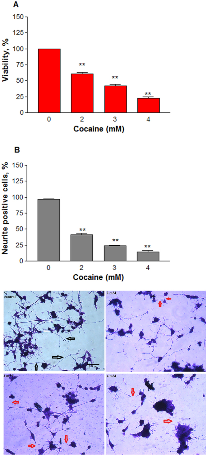
Cytotoxic effect of cocaine at higher doses. Differentiated cells in phenol-red free medium were treated with PBS (control) or cocaine for 48 h. Cell viability (A) was assessed (n = 12, **P = 0.001) by the crystal violet dye uptake method; neuronal extensions greater than 2-body diameter in length of 100 cells per well at four randomly chosen field areas were measured (n = 8, **P = 0.001) and quantified (B); the crystal violet stained cells were photographed under an inverted phase contrast 1X-70 Olympus microscope with 20x objective (C), showing neurite connections with black arrows or disconnected neurites upon cocaine treatments with red arrows. Data are expressed as mean ± SEM, significant compared to the PBS controls. Scale bar 0.06 cm.
