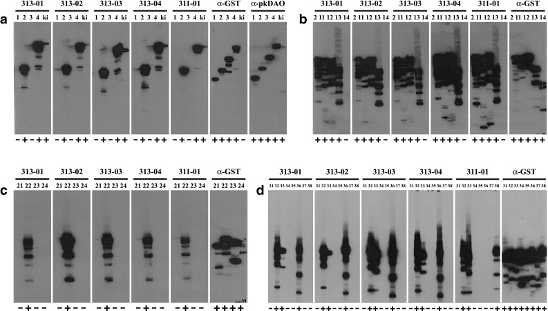
Fig. 2.

Antibody binding to DAO fragments. Antibody binding was tested using filter strips of DAO fragments expressed from pGEX-huDAO01-04 used for immunization (a), of DAO fragments expressed from pGEX-huDAO11-14 with C-terminal deletions of pGEX-huDAO02 (b), of DAO fragments expressed from pGEX-huDAO21-24 with HaeIII fragments of pGEX-huDAO02 (c), or of DAO fragments expressed from pGEX-huDAO31-38 obtained by subcloning of pGEX-huDAO22 (d). Filter strips containing approximately 5 µg cell lysate protein separated on a 12.5% SDS polyacrylamide gel were incubated with the mouse monoclonal antibodies HYB313-01/-02/-03/-04, HYB311-01 (diluted 1:7500–1:20,000 in TBSTM), the anti-GST antibody HYB374-01 (1:1500), or the rabbit polyclonal antibody α-pkDAO (1:10,000) made against porcine kidney DAO, respectively. Filter strips were then incubated with horseradish peroxidase-conjugated anti-mouse immunoglobulins (1:1500 in TBSTM) or anti-rabbit immunoglobulins (1:5000), respectively, followed by ECL substrate, and exposure to film for 0.25–10 min. Lane numbers 1–4, 11–14, 21–24, and 31–38 correspond to expression constructs pGEX-huDAO01-04, pGEX-huDAO11-14, pGEX-huDAO21-24, and pGEX-huDAO31-38, respectively, and ki to a human kidney lysate. Binding is indicated by + and − signs below each lane. Exact positions of the bands on parallel lanes vary slightly because filter strips from different individual blots were used for this experiment. Besides the major band of the respective full-length fusion protein, a variable number of smaller bands is visible in most lanes due to production of partial products in the bacterial expression system
