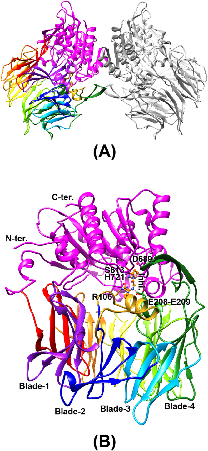
Figure 1.

Three-dimensional structure of PmDAP IV. The catalytic domain is coloured in magenta. The β-propeller domain is coloured from purple (blade-1) to red (blade-8). A long insertion containing Arg106 located between blade-1 and blade-2, which was disordered in the ligand-free form, is shown in gold. (A) Dimeric structure of PmDAP IV. (B) PmDAP IV subunit. The catalytic triad “Ser613, Asp689, and His721”, the double-Glu motif “Glu208-Glu209”, Arg106 located between blade-1 and blade-2, a bound inhibitor molecule, and blades 1 to 4 are labelled.
