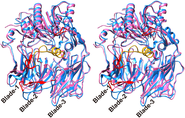
Figure 4.
Wall-eyed stereo view showing a conformational difference of PmDAP IV. Ligand-free (cyan) and dipeptide-bound (magenta) forms of PmDAP IV are shown. Least-square fitting was performed with respect to all Cα atoms of each molecule. Cα-Cα deviations larger than 5 Å are shown in red for the dipeptide-bound form. A long insertion containing Arg106 located between blade-1 and blade-2, which was disordered in the ligand-free form, is shown in gold.

