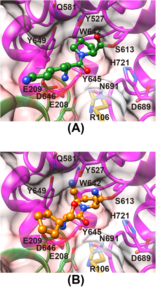
Figure 5.

The mode of peptide/inhibitor binding in PmDAP IV. Residues belonging to the catalytic domain and blade 4 of the β-propeller domain are coloured magenta and green, respectively. The side chain of Arg106 is shown in gold. The surfaces are coloured in blue and red by a coulombic surface colouring to indicate positive and negative charges, respectively, from 30 kT to -30 kT. (A) Lys-Pro (green) complex. (B) Inhibitor-1c (orange) complex.
