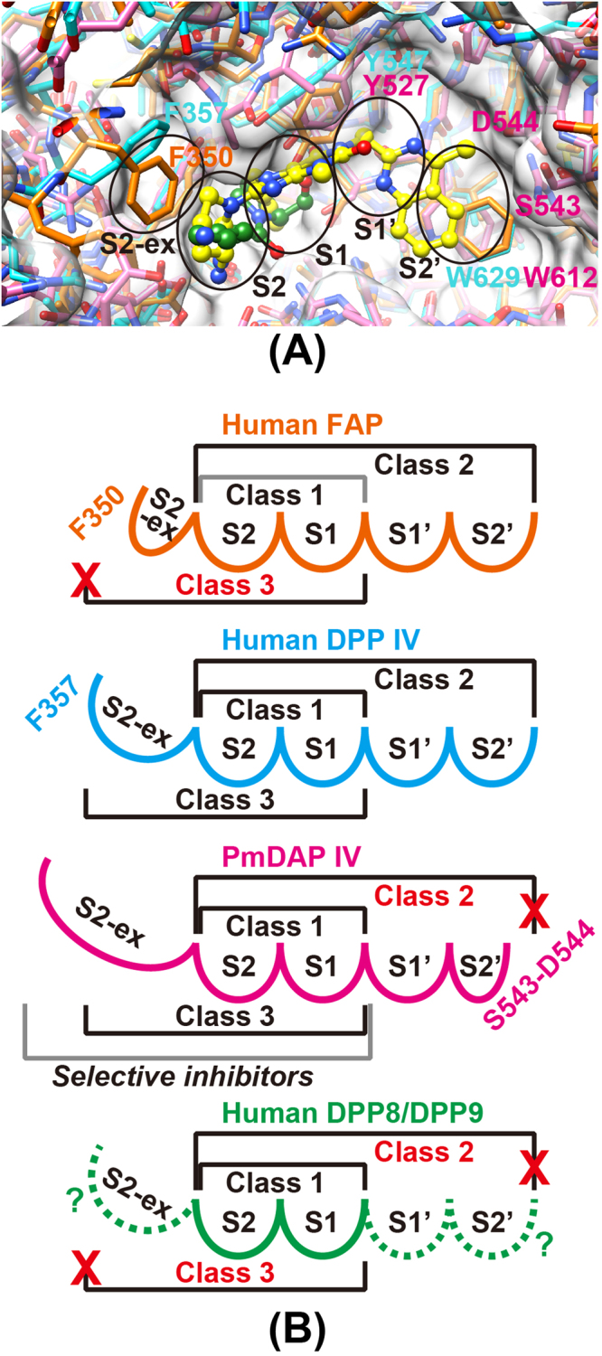
Figure 8.

Active site cleft of PmDAP IV and human DPP IV-family enzymes. (A) Superposition of the active site of the Lys-Pro (green) complex of PmDAP IV (magenta) and those of the linagliptin (yellow) complex of human DPP IV (cyan) and ligand-free human FAP (orange). Subsites are indicated by ellipsoids. (B) A schematic figure showing the gliptin-binding sites of PmDAP IV and DPP IV-family enzymes. The diagrams of FAP, DPP IV, and PmDAP IV are drawn based on their crystal structure, whereas that of DPP8/DPP9 is a conceptual diagram.
