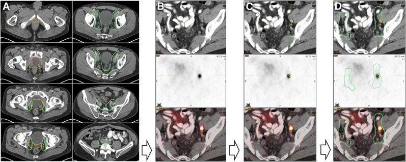
FIGURE 1.
Study methodology. (A) Experienced radiation oncologist masked to PET findings contoured RTOG CTVs onto CT dataset of PET/CT scan for all 270 patients (prostate bed CTV in orange and pelvic LN CTV in green). (B) All 68Ga-PSMA-11 PET/CT images were analyzed by an experienced nuclear medicine physician. (C) PSMA-11–positive lesions were contoured in yellow on CT images. (D) Consensus CTVs were coregistered with 68Ga-PSMA-11 PET/CT images and PSMA-11–positive lesion contours (yellow) to assess, for each patient, whether PSMA-11–positive lesions were localized inside or outside consensus CTVs.

