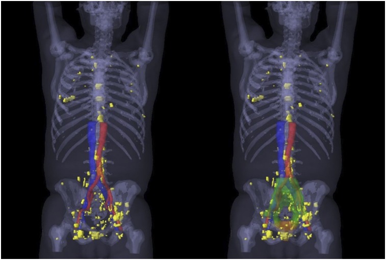
FIGURE 2.
A 3-dimensional map of the PSMA-11–positive lesions (yellow) of all 52 patients with recurrence outside consensus CTVs (23 patients with recurrence outside only and 29 patients with recurrence outside and inside consensus CTVs), created by rigid registration of each patient’s CT image to template patient’s CT image, followed by transfer of each PSMA-11–positive lesion contour to template patient CT image (MIM, version 6.7.5; MIM Software Inc.). On right side, 3-dimensional prostate bed consensus CTV is shown in orange and 3-dimensional pelvic LN consensus CTV in green.

