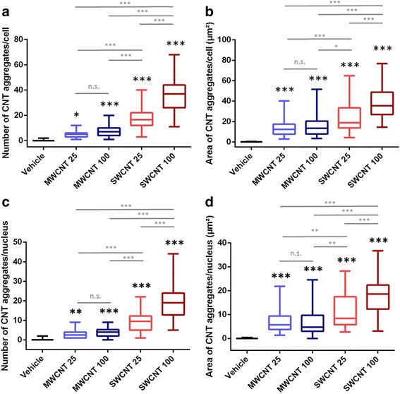
Fig. 3.

Quantitative analysis of the cellular and nuclear deposition of MWCNTs and SWCNTs. a Number of CNT aggregates per cell; b total area of CNT aggregates per cell (μm2); c number of CNT aggregates per nucleus, d total area of CNT aggregates per nucleus (μm2). All conditions were significantly different from the control condition (vehicle) and from each other (one way ANOVA, Tukey multiple comparison) except for the different doses of MWCNTs [not significant (n.s.)]. The box plots represent median and quartiles, and the whiskers represent the 1.5 interquartile range of the lower and upper quartile (N = 50 for each condition). 25 and 100 represents exposure concentrations as μg/ml
