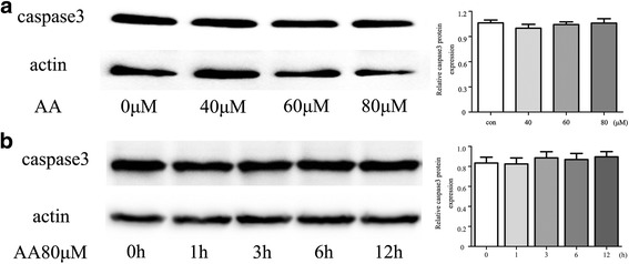
Fig. 2.

Caspase-3 expression in RAW264.7 cells. a RAW264.7 cells were pre-treated with AA for 12 h at the indicated concentrations. b RAW264.7 cells were pre-treated with 80 μM AA for the indicated length of time. Total protein was immunoblotted with Caspase-3 antibody. The figure containes the densitometric quantification of relative protein expression
