
Fig. 3.
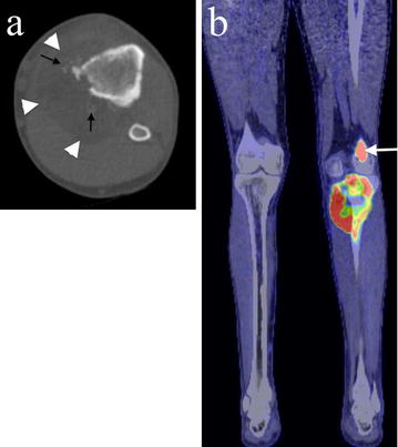
Axial CT and PET/CT imaging of the proximal tibial lesion. a Axial CT imaging shows medial cortical destruction and a large soft tissue mass containing areas of fat attenuation (white arrowheads) and ossification (black arrows). b A coronal fused PET/CT image shows there is marked FDG uptake in the proximal tibial tumour as well as in a lateral distal femoral metastasis (white arrow). No other lipomatous lesion or tumour is present