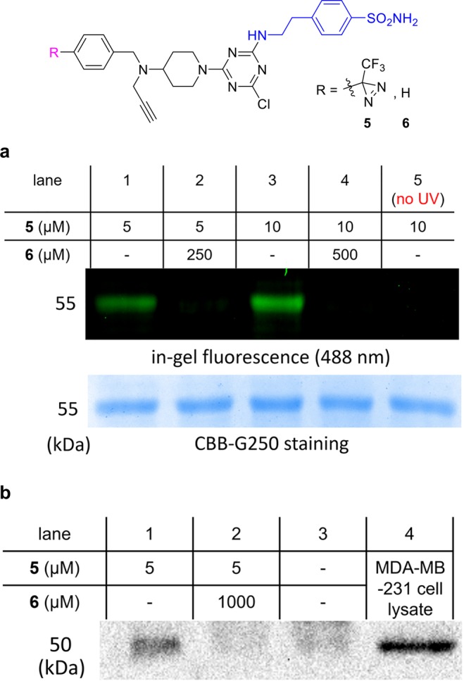
Figure 2.

(a) Experiments with isolated protein. Photoaffinity labeling of recombinant CAIX after 30 min illumination with excess ligand 5 and click with azide-fluor-488; the top gel (black background) shows the fluorescence at 488 nm, and the one below depicts protein staining with Coomassie Brilliant Blue (CBB) G250. Competition with 6, which lacks a diazirine, suppresses labeling with 5 (lanes 2 and 4). (b) Experiments with MDA-MB-231 cell lysates. MDA-MB-231 cell lysate was treated with PAL probe 5 under 365 nm illumination for 30 min and then clicked with biotin-azide. NeutrAvidin agarose was used to pull down the biotinylated proteins, and the material captured was run on a SDS-PAGE gel. Lane 1 shows staining of CAIX on the Western blot image with a CAIX Ab, lane 2 shows sample pretreated with blocking ligand 6, lane 3 shows no ligands, and lane 4 depicts the lysate without PAL.
