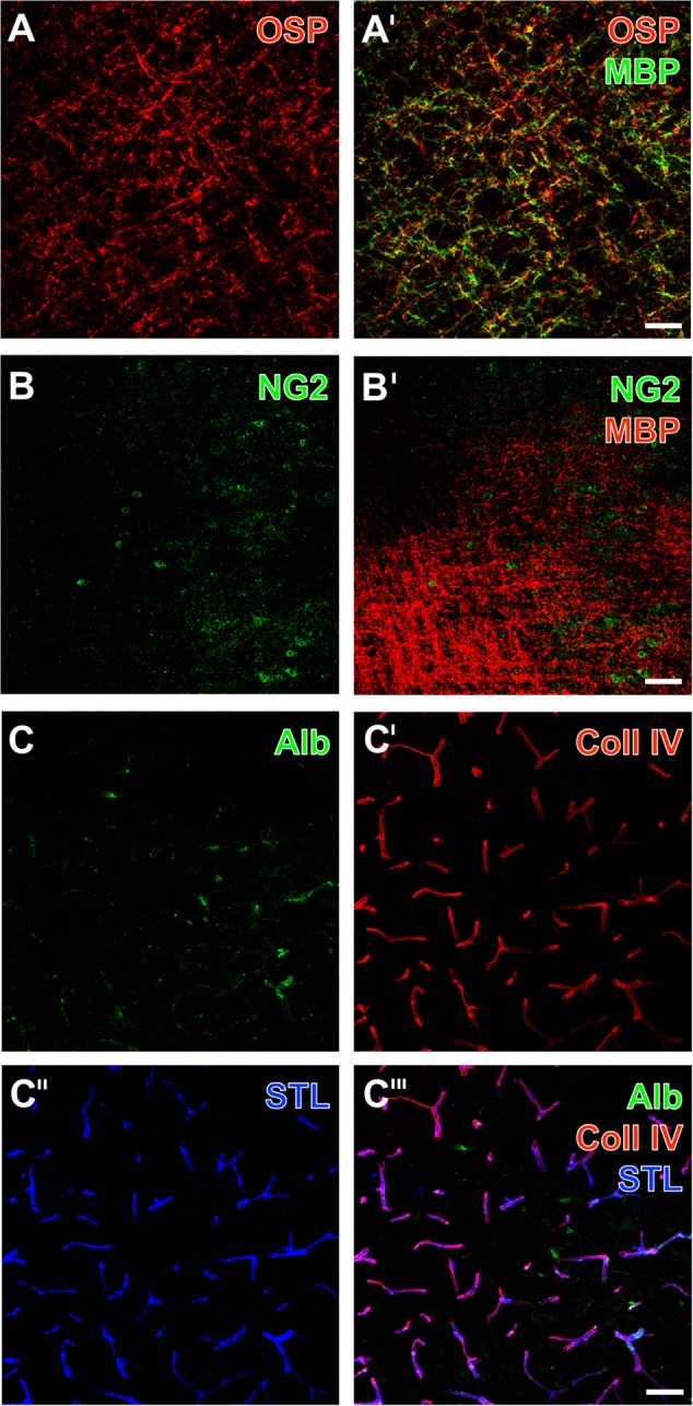
FIGURE 1.

Representative ischemia-related alterations of oligodendrocyte structures and the vasculature in the striatum 1 day after experimental focal cerebral ischemia in 3-month-old (C) as well as in 12-month old mice (A,B), captured by laser scanning microscopy. Concomitant fluorescence staining of the oligodendrocyte-specific protein (OSP, A) and the myelin basic protein (MBP, A′) as myelin-associated markers exhibited a dense network of dotted and strand-like structures, which appeared visually enhanced due to ischemia. Immunoreactivity of the neuron-glia antigen-2 (NG2) was nearly absent in these tissues (B), while simultaneous detection with MBP clearly showed enhancement of the MBP in the same region (B′). Endogenous serum albumin was found in ischemia-affected areas (C) indicating impaired blood – brain barrier integrity, while visualization of the vasculature by collagen IV (Coll IV)-immunostaining (C′) and detection of Solanum tuberosum lectin (STL)-binding sites (C″) appeared robustly with a wide range of overlapping vascular structures (C″′). Scale bars: A′ (also valid for A) = 50 μm, B′ (also valid for B) = 50 μm, C″′ (also valid for C–C″) = 25 μm.
