
FIGURE 2.
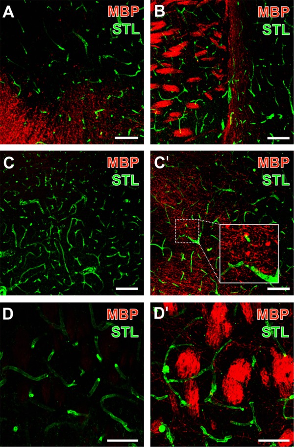
Representative micrographs from double fluorescence labeling of myelin basic protein (MBP) and endothelial STL-binding sites 1 day after focal ischemia in the affected striatum of 3-month-old (B–D) as well as 12-month-old mice (A), visualized by confocal laser-scanning microscopy. MBP-immunodetection revealed an increased signal toward the ischemic zone (upper part of A, left part of B). Inter-hemispheric comparison demonstrated the nearly absent immunoreactivity of MBP (C) while vessels were clearly visible as detected by STL (C) on the contralateral, non-affected hemisphere. The same striatal region on the ischemia-affected hemisphere exhibited a dense network of MBP-positive structures (C′) in close regional association to the vasculature (inset in C′). At higher magnifications, densely packed fiber-like structures of MBP were regularly observed in the ischemia-affected striatum (D′), while the contralateral striatum was devoid of these MBP-positive structures (D). Scale bars: (A) = 75 μm, (B) = 100 μm, (C,C′) = 100 μm, (D,D′) = 50 μm.