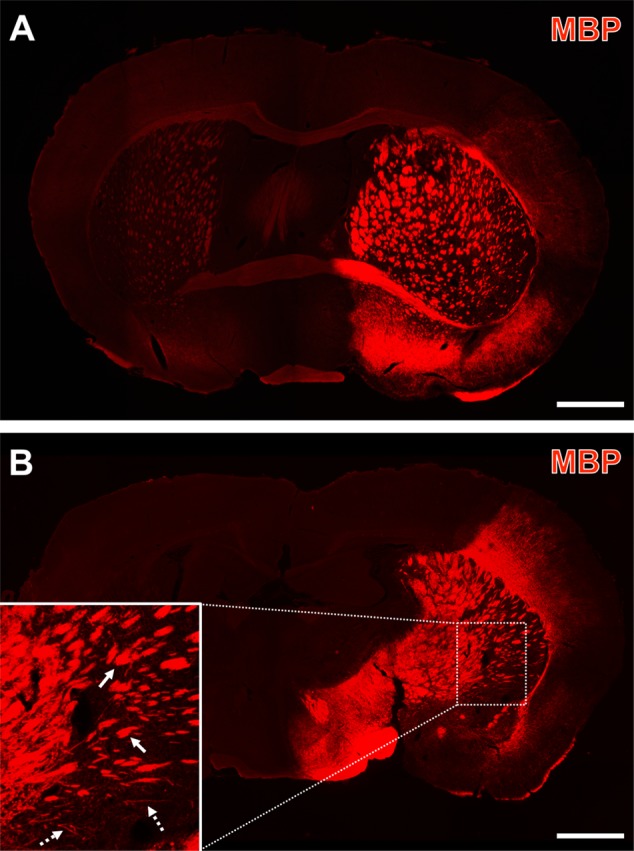
FIGURE 3.

Representative forebrain overview scans from immunolabeled myelin basic protein (MBP) 1 day after focal cerebral ischemia in 3-month-old (A) and a 12-month-old mice (B). The enhanced MBP-immunosignal captured the overall territory of the middle cerebral artery (i.e., the ischemia-affected striatum, some thalamic regions and lateral parts of the neocortex), which was occluded by a filament-based model of focal cerebral ischemia. Higher magnification (inset in B) indicated dense packages of MBP-immunopositive structures (arrows with plain lines) predominantly in subcortical regions and – toward cortical areas – groups of MBP-positive, mostly parallel arranged strands (arrows with dashed lines). Scale bars: (A,B) = 1 mm.
