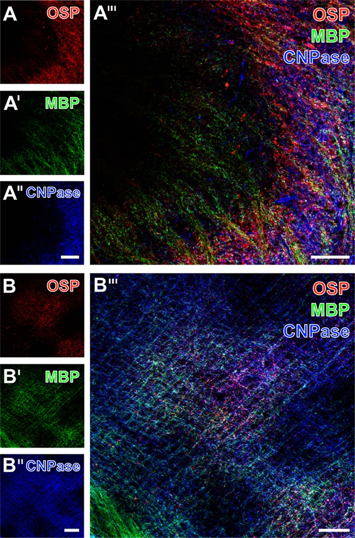
FIGURE 6.

Representative micrographs of a 12-month-old mice 1 day after focal cerebral ischemia demonstrating triple fluorescence labeling of the oligodendrocyte-specific protein (OSP), myelin basic protein (MBP), and 2′,3′-cyclic nucleotide-3′-phosphodiesterase (CNPase). Immunoreactivities for OSP and MBP were enhanced in the ischemic striatum (right part in A and A′) and the lateral neocortex (B,B′). In a similar manner, the immunosignal of CNPase appeared to be enhanced in the ischemic striatum (right part in A″) too, which became even clearer when merging the staining pattern (A″′). Further, an enhanced CNPase-immunoreactivity appeared in the ischemia-affected neocortex except for upper neocortical layers (upper left corner in B″), while overlaying structures were predominantly identified in the affected neocortex (middle part of B″′). Scale bars: A″ (also valid for A and A′) = 100 μm, A″′ = 50 μm, B″ (also valid for B and B′) = 100 μm, B″′ = 50 μm.
