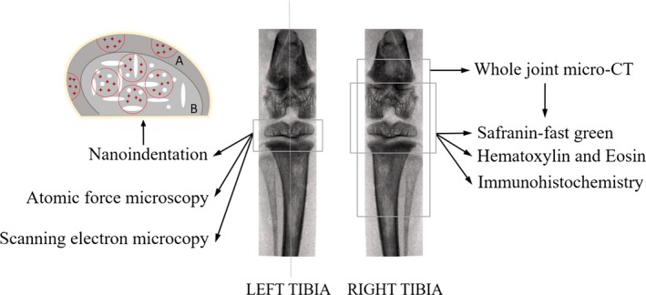
Figure 1.

Synopsis of site‐specific measurements performed in the tibial articular cartilage (AC) and subchondral bone regions, and sketch map of indentation areas and sites in sagittal plane of knee joints. Considering the structural differences between subchondral plate (SP) and trabecular bone, the outer edge of the sample was set as a reference. According to the sample size, three–five semi‐circles with 0.5 mm diameter were selected as indentation areas of SP (A), and three–five circles with 0.8 mm diameter were selected as indentation areas of trabecular bone (B). The red crosses in the sketch map were indentation sites, and three–five indentation sites were selected in each indentation area. Indentation sites were dispersedly distributed in each indentation area to ensure that locations of bone tissue can be selected as much as possible for the nanoindentation test; besides, the indentation sites should be far away from the edge in order to diminish the influence of embedding on the results.
