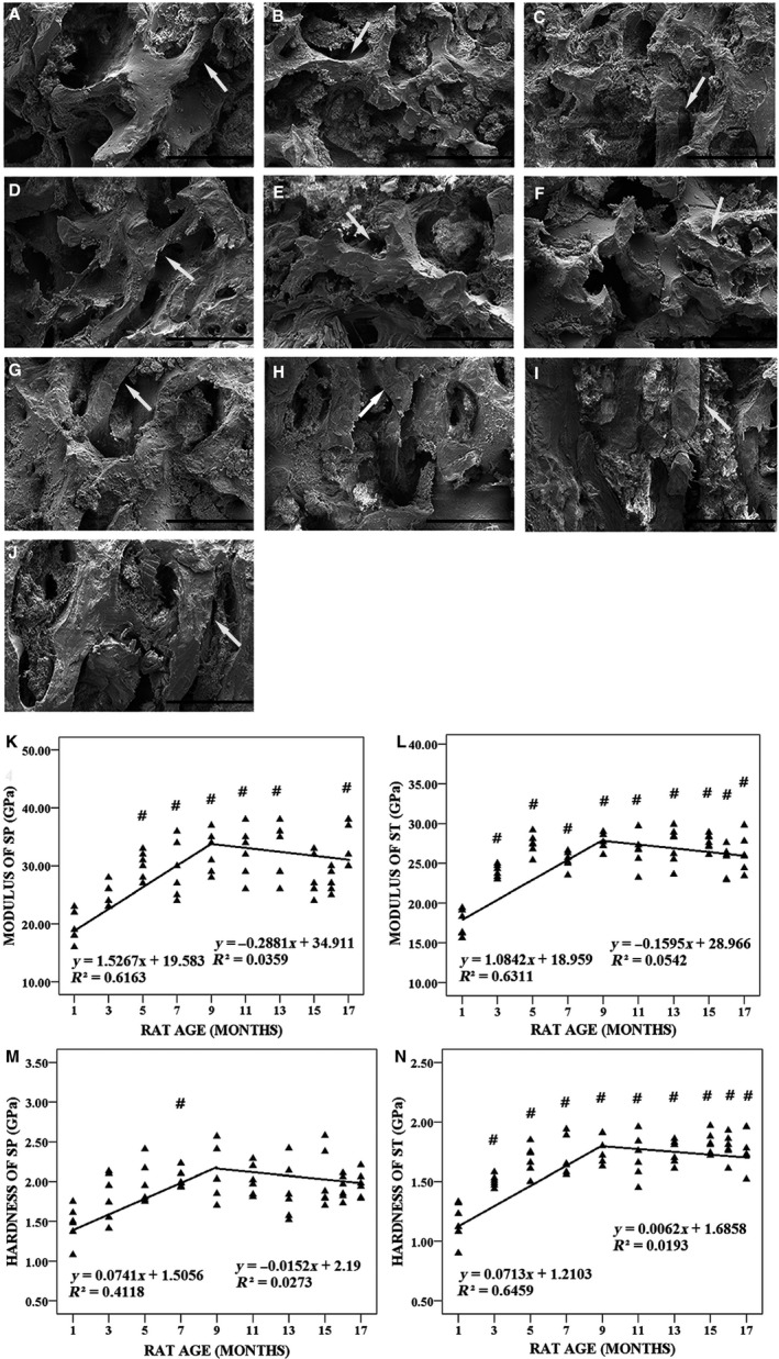
Figure 7.

Typical microarchitecture features of the surfaces of subchondral trabecular bone (ST) in tibial plateau from 1‐, 3‐, 5‐, 7‐, 9‐, 11‐, 13‐, 15‐, 16‐ and 17‐month‐old rats detected by scanning electron microscopy (SEM) and indentation modulus and hardness of subchondral plate (SP) and ST in tibial plateau measured by nanoindentation test (n = 100). (A–J) Typical SEM images of the surfaces of ST in tibial plateau from 1‐, 3‐, 5‐, 7‐, 9‐, 11‐, 13‐, 15‐, 16‐ and 17‐month‐old rats, respectively. Bars represent a length of 300 μm. The white arrows indicate age‐related changes of ST. (K) The indentation modulus of SP in tibial plateau was plotted with animal age. (L) The indentation modulus of ST in tibial plateau was plotted with animal age. (M) The hardness of SP in tibial plateau was plotted with animal age. (N) The hardness of ST in tibial plateau was plotted with animal age. # P < 0.05, compared with 1‐month group.
