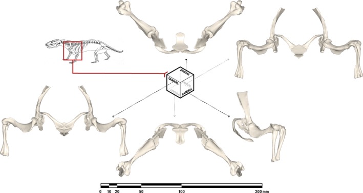
Figure 3.

Orthographic views of the pectoral limb of Massetognathus pascuali in an anatomically neutral reference pose (not in vivo posture), with all joints rotated to the centers of their measured ranges of motion. The bones depicted comprise the bilateral scapulocoracoids, humeri, ulnae, and radii, as well as the median interclavicle. Line drawing of M. pascuali adapted from Fig. 9 in Jenkins (1970b).
