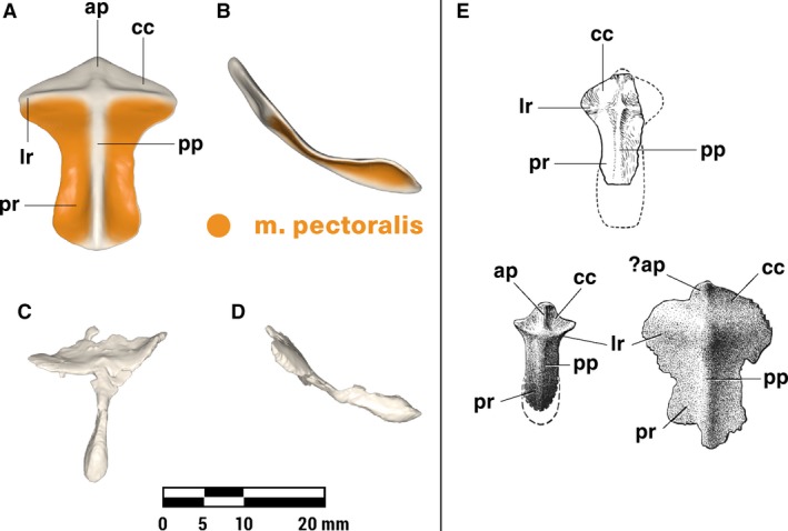
Figure 5.

Repaired (A, B) and original (C, D) interclavicle of Massetognathus pascuali in ventral (A, C) and lateral (B, D) views, with reconstructed muscle origins/insertions. Reference images (E) adapted from Jenkins (1970b) (top, M. pascuali) and Jenkins (1971a) (bottom left Thrinaxodon, bottom right unidentified cynodont.) Abbreviations follow Jenkins (1971a): ap, anterior ridge; cc, concavity for clavicle articulation; lr, lateral ridge; pp, posterior ridge; pr, posterior ramus. Reconstructed muscles are listed in legend.
