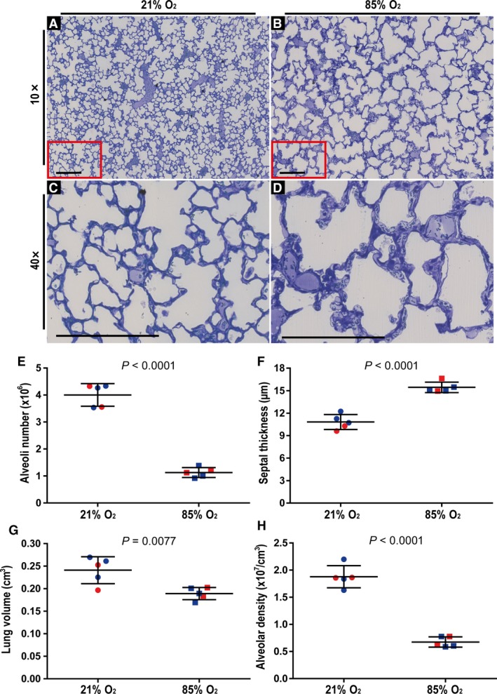
Figure 1.

Validation of the impact of hyperoxia exposure on post‐natal lung alveolarisation in an experimental mouse model of bronchopulmonary dysplasia. Visual inspection of histology sections from mice exposed to (A) 21% O2 revealed normal lung architecture, whereas (B) a reduction in the number and an increase in the size of the alveoli was clearly evident in lungs from mice exposed to 85% O2 for the first 14 days of post‐natal life. Similarly, under higher magnification (C,D), an increase in the arithmetic mean septal thickness comparing lung sections from (C) 21% O2‐exposed mouse pups and (D) 85% O2‐exposed mouse pups, was clearly evident. Sections represent the same trends observed in another four mice in each experimental group. (C,D) Higher‐magnification images of the regions demarcated by the red box in (A) and (B), respectively. Scale bar: 200 μm. The (E) total number of alveoli in the lung, (F) arithmetic mean septal thickness, (G) lung volume, and (H) alveolar density were quantified by stereology analyses and lung volume determination using Cavalieri's principle. Male animals are indicated by blue symbols, and female animals by red symbols. Data reflect mean ± SD (n = 5 animals per experimental group). Comparisons were made by unpaired Student's t‐test.
