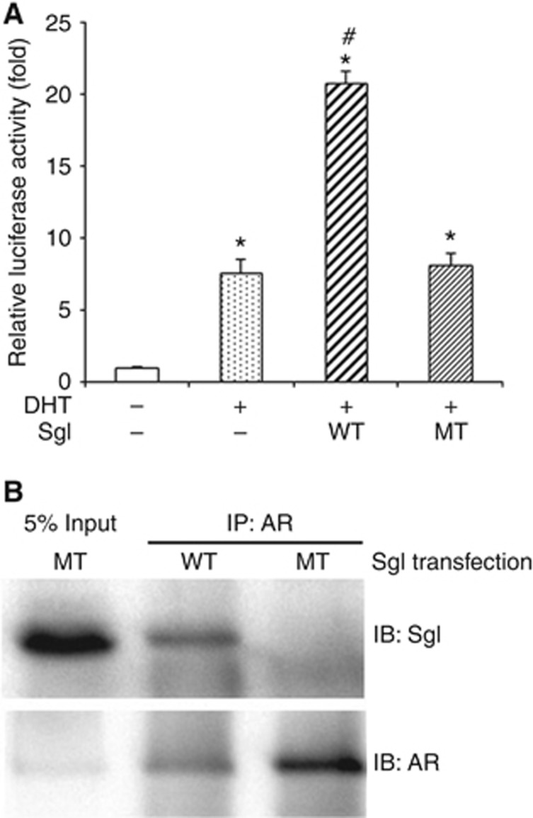
Figure 2.
The effects of wild-type (WT) vs mutant (MT) SgI on AR activity in prostate cancer cells. (A) PC3 cells were co-transfected with pSG5-AR (5 ng), pSG5 or pSG5-SgI (250 ng), MMTV-luciferase (250 ng), and pRL-TK (2.5 ng), and cultured in phenol red-free medium supplemented with 5% charcoal-stripped FBS along with zinc (100 μM) and either mock (ethanol) or 10 nM DHT for 24 h. The luciferase activity is presented relative to that of mock treatment (first lane; set as one-fold). Each value represents the mean+s.d. of three independent determinations. *P<0.05 (vs control cells with mock treatment). #P<0.05 (vs control cells with DHT treatment). (B) Co-precipitation of AR and SgI. Cell lysates (500 μg) from PC3 transfected with pSG5-AR (7 μg) and pSG5-SgI (7 μg) and cultured in medium supplemented with 10% normal FBS in the presence of 300 μM zinc were incubated with an anti-AR polyclonal antibody (2 μg). Input protein (25 μg of total lysates) and the precipitated proteins were resolved on a 10% SDS–PAGE and blotted with an anti-SgI or anti-AR antibody.

