Figure 1.
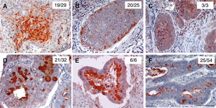
The presence of Td-CTLP in orodigestive tumour tissues. Immunohistochemical stainings of the Td-CTLP in tongue (A), tonsillar (B), oesophageal (C), gastric (D), pancreatic (E), and colon cancer (F) tissues. Number of positive tissue samples per total sample size for each tumour type shown. 3-amino-9-ethylcarbazole (AEC) was used as chromogen (red). All red-stained areas on each tissue section indicate specific detection of Td-CTLP. Scale bar is 100 μm, relevant for all panels.

