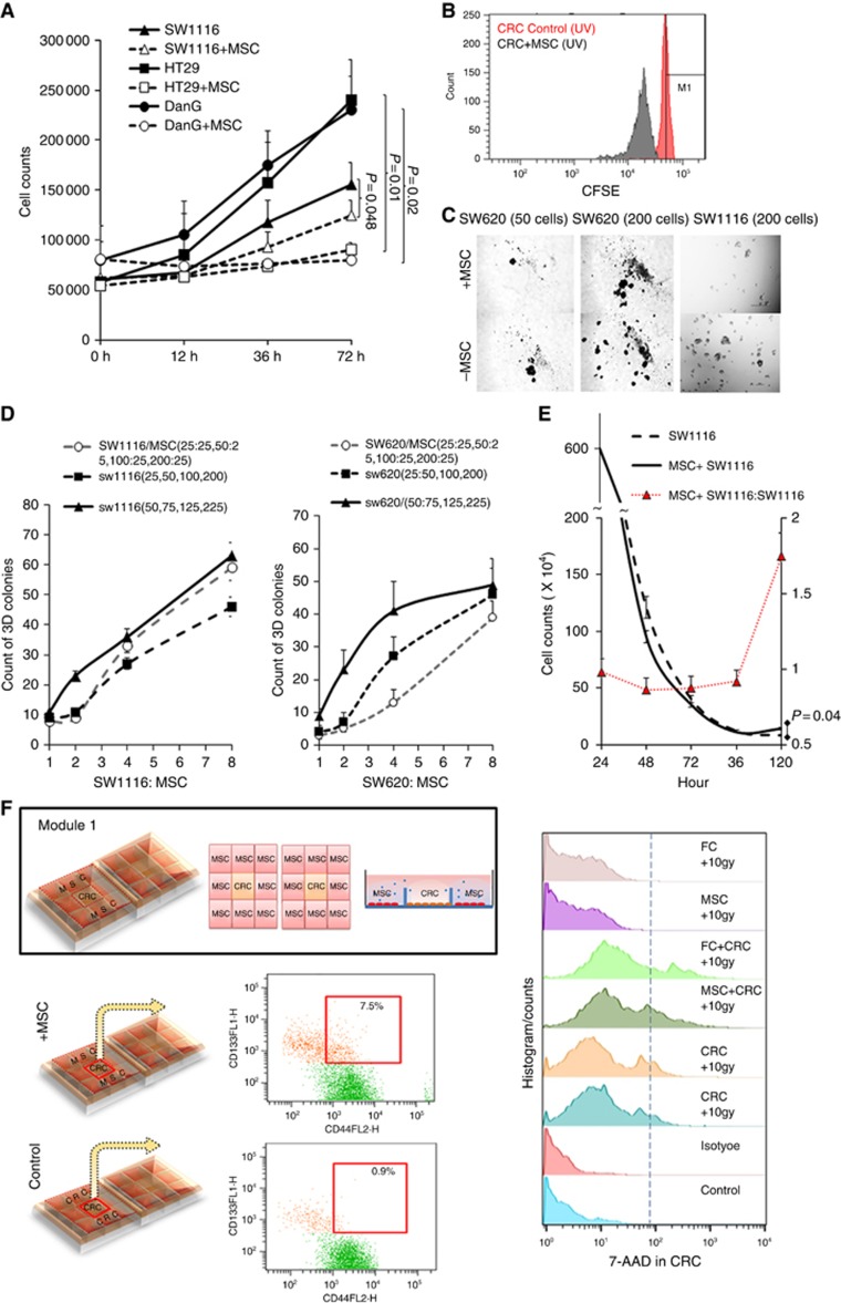
Figure 2.
Co-culture system showed attenuate proliferation and viability under UV irradiation. (A) This is a representative figure of proliferation experiment. In total, 4 × 10⁴ SW1116, HT29 or DanG cells were seeded with or without (control group) 1 × 10⁴ MSCs in 12-well plate. In total, 10 J cm−2 irradiation was performed for 1 h and last for 6 h and total cell numbers in each well were counted at 12 h, 36 h, and 72 h respectively. (B) CSFE assay of control and co-cultivation group. (C, D) 25, 50, 100, or 200 SW1116, SW620 cells were seeded in ultra-low attachment 24-well plates, respectively (control), or following by additional 25 bone marrow-derived mesenchymal stromal cells seeding in each well. Twenty-five more SW1116 cells were also seeded instead of 25 MSCs as blank control. After 10 J cm−2 irradiation 1 h per 8 h for 8 days, cell colonies were counted on the 9th day. Independent experiments were repeated 2∼3 times. Mean value were represented. (E) 6 × 10⁶ SW1116 cells were seeded in co-culture system (Supplementary Figure 2B), with or without 106 MSC cells seeding in the inserted well. 10 J cm−2 irradiation was performed for 1 h and last for 8 h and total cell numbers in each well were counted at 24 h, 48 h, 72 h, 96 h, and 120 h, respectively. The colorectal cancer cells decreased more rapidly in the co-culture group than control group (SW1116 only) at the beginning (0∼96 h) after irradiation, however, turned to be slower and stayed stable after 96 h. (F) Co-culture model for MSCs and CRC. The μ–Slide 2 × 9 well harbours two arrays of 3 × 3 square fields where cells can be cultivated independently within the small square or share the same growth medium within the total 3 × 3 square fields. After co-cultivation for 48 h, flow cytometry showed more CD133+CD44+ colorectal cancer stem cell-like cells than the control group. However, there were also more 7-AAD+dead cells compared with the control group. *P<0.05.

