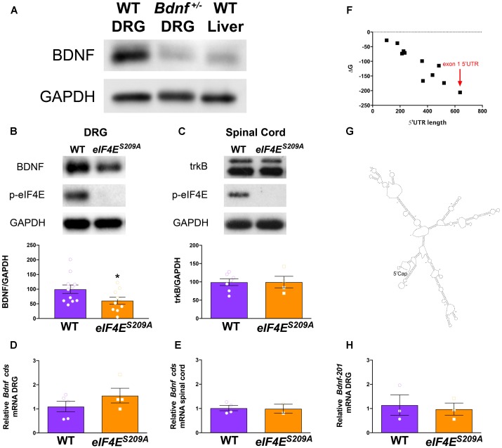
FIGURE 1.
Decreased Bdnf mRNA translation in eIF4ES209A mouse DRG. BDNF antibody was verified by immunoblotting against Bdnf+/- DRGs and WT liver showing reduced levels of BDNF protein compared to WT DRGs (A). eIF4ES209A mouse DRGs (B, n ≥ 4, t = 3.238, df = 7, ∗p = 0.0143, t-test) showed lower levels of BDNF protein expression compared to WT (n ≥ 4, ∗p < 0.05, t-test) but equal levels of trkB expression (C) in the spinal cord (n ≥ 5, t-test). (D,F) While BDNF protein levels were lower in eIF4ES209A DRG compared to WT DRG, total-Bdnf mRNA levels were equal in DRG (D) and in spinal cord (E, n ≥ 4, t-test). (F) Delta G (ΔG) free energy map of Bdnf transcript variants plotted by 5′ UTR length. Bdnf-201 transcript is shown by the red arrow. (G) Structure of Bdnf exon 1 5′ UTR as predicted by mfold: http://unafold.rna.albany.edu/?q=mfold. (H) Bdnf-201 mRNA expression was equal in DRG between eIF4ES209A and WT DRG (n ≥ 4, t-test).

