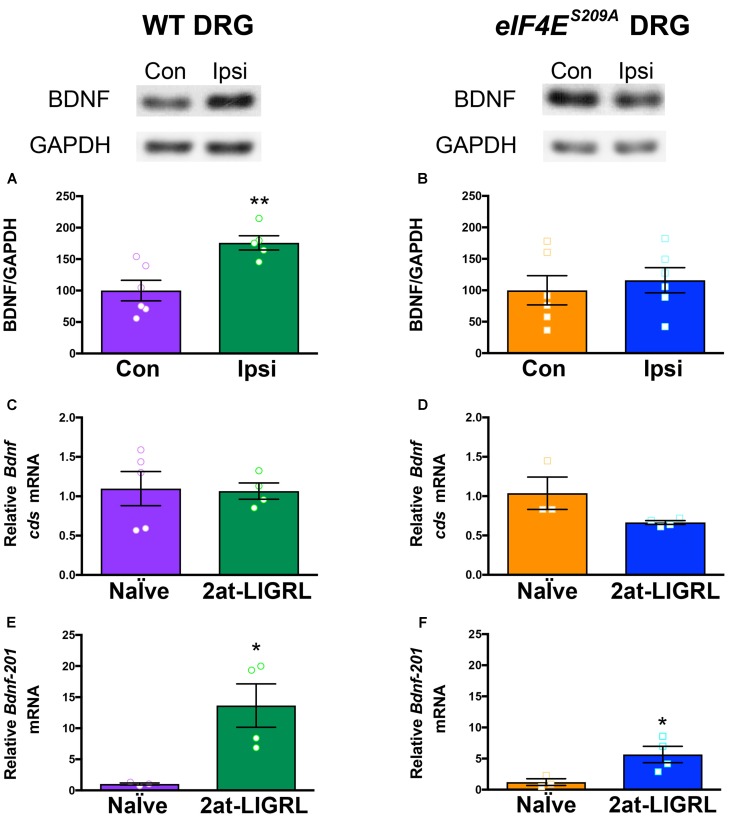
FIGURE 3.
BDNF protein is increased in WT but not eIF4ES209A DRGs after PAR2 activation. (A) Western blot analysis shows an increase in BDNF protein standardized to GAPDH in WT DRGs (L4-L6) ipsilateral (IPSI) to 2at-LIGRL intraplantar (i.pl.) injection compared to contralateral (CON) (n ≥ 5, t = 3.662, df = 9, ∗∗p = 0.0052, t-test). Protein and mRNA extraction was done 24 h following i.pl. injection. (B) eIF4ES209A DRG (L4-L6) showed no differences in BDNF protein levels between IPSI and CON (n = 6, p > 0.05, t-test). mRNA levels of Bdnf-pan and the Bdnf-201 isoform were analyzed using qPCR. (C,D) While Bdnf-pan mRNA levels after 2at-LIGRL injection were not changed in DRGs from both genotypes (n ≥ 3, t-test). (E,F) Bdnf-201 mRNA levels were significantly increased in both WT and eIF4ES209A DRGs (WT: n ≥ 3, t = 3.055, df = 5, ∗p = 0.0283, t-test; eIF4ES209A: n ≥ 3, t = 2.756, df = 5, ∗p = 0.04, t-test).

