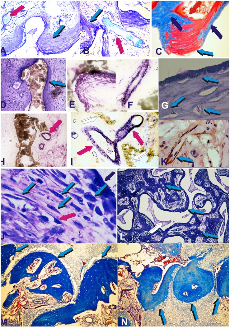
Figure 1.

Self-inducing geometric cues and the induction of tissue patterning, morphogenesis with the final induction of bone formation as initiated by the geometric concavities of the coral-derived calcium phosphate based macroporous bioreactors. Coral-derived macroporous bioreactors, 20 mm in height and 11 mm in diameter, were implanted in heterotopic intramuscular rectus abdominis sites in a series of non-human primate Chacma baboon Papio ursinus. Generated tissues were harvested on day 30, 60, and 90 after heterotopic implantation (Ripamonti, 1990, 1991, 1996; Ripamonti et al., 1993, 2009, 2010; van Eeden and Ripamonti, 1994). Harvested tissues were processed for decalcified and undecalcified histological analyses. (A–D) Tissue patterning and the induction of mesenchymal tissue condensations on day 30 (day 90 in C) at the hydroxyapatite interface (light blue arrows) with capillary invasion (magenta arrows). Alkaline phosphatase staining and activity within both mesenchymal condensations (on day 60) (E) and capillary sprouting and invasion (on day 30) (F). (G) Patterning and further morphogenesis of collagenous condensations at the hydroxyapatite interface with the development of osteoblast-like cells within the differentiating and remodeling condensations on day 30 (light blue arrows). (H,I) Further alkaline phosphatase staining (magenta arrows) of invading capillaries on day 30. Alkaline phosphate stains intensely within the multiple cellular layers of the sprouting branching capillaries in close contact with the hydroxyapatite substratum. (K) Macroporous construct harvested on day 60 shows laminin immunolocalization (light blue arrow) within invading capillaries penetrating the macroporous spaces. Type IV collagen and laminin' amino acid motifs bind both angiogenic and bone morphogenetic proteins sequences which may be released during tissue induction and morphogenesis to initiate the induction of bone formation (Ripamonti, 2006, 2010 for reviews) Note the intimacy of laminin immunolocalization with large hyperchromatic endothelial cells, possibly preparing to migrate out of the vascular compartment. (J) On day 60, there is a continuous flow of responding mesenchymal cells (light blue arrows) moving from the vascular Trueta's angiogenic vessels (magenta arrow) to the osteoblastic/osteogenetic differentiation site (dark blue arrow). The digital image in (J) depicts the critical differentiating role of the nanopatterned geometric substratum on the induction of cellular differentiation, osteoblast synthesis and the induction of bone formation directly attached to the self-inducing nanopatterned calcium phosphate-based macroporous bioreactor (Ripamonti et al., 1993). (L) Bone morphogenesis later develops on day 90 within the macroporous spaces of the coral-derived bioreactors, with woven bone (light blue arrows in L) with woven bone initiating within concavities of the substratum. (M,N) Bone induction and remodeling of the newly formed bone within concavities (light blue arrows) on day 90 after rectus abdominis implantation. Decalcified and undecalcified paraffin wax and Historesin sections cut at 3 to 6 μm and stained with toluidine blue in 70% ethanol or (C) stained free-floating with modified Goldner's trichrome stain (C). Decalcified sections (M,N) also stained with modified Goldner's trichrome stain.
