
Figure 4.
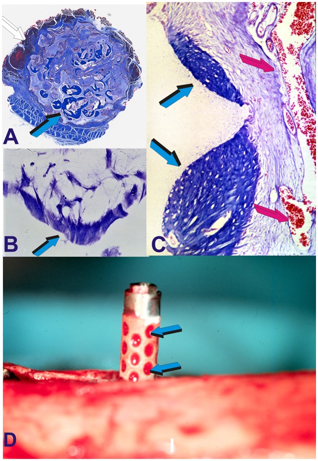
Molecular, morphological and biological conceptualization of the role of the concavity to construct intrinsically osteoinductive hydroxyapatite-coated titanium implants for the spontaneous induction of bone formation. (A) Substantial induction of bone formation (light blue arrow) by highly sintered highly crystalline hydroxyapatite bioreactors implanted in the rectus abdominis muscle of the Chacma baboon Papio ursinus (Ripamonti et al., 1999). (B) Alignment and orientation of MC 3T3-E1 pre-osteoblastic cells within a concavity (light blue arrow) of the coral-derived bioreactor in vitro (Ripamonti et al., 2012a). (C) Digital image of a macroporous sintered hydroxyapatite resembling the tread of an implant implanted heterotopically yet spontaneously inducing bone (light blue arrows) within the concavities of the bioreactor in close relationship with invading capillaries (magenta arrows). (D) The “concavity motif,” the “concavity: the shape of life” (Ripamonti, 2006; Ripamonti et al., 2012a) is then re-assembled in a titanium construct prepared with concavities along the substratum later coated by highly crystalline plasma sprayed sintered hydroxyapatite onto the prepared titanium surface (Ripamonti et al., 2012a). Light blue arrows (D) indicate the adsorption within concavities of plasma products during the surgical implantation in orthotopic mandibular and tibial sites (Ripamonti et al., 2012a).