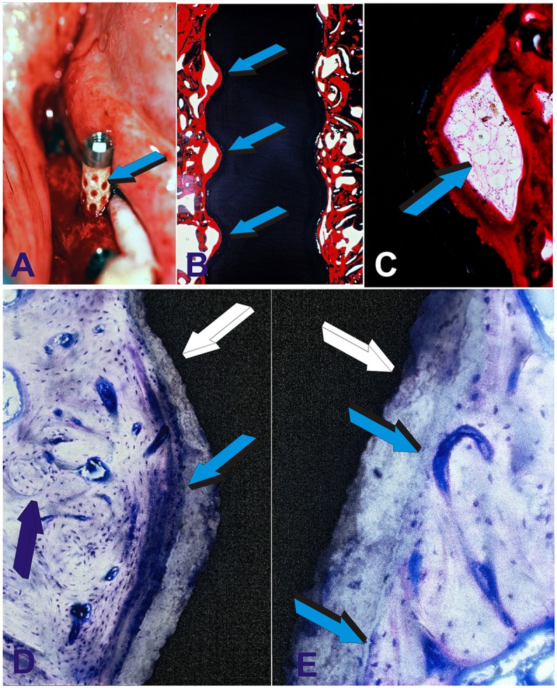
Figure 8.

Preclinical testing of the geometric self-inductive constructs in edentulous mandibular ridges of adult Chacma baboons Papio ursinus. (A) Highly crystalline hydroxyapatite coated titanium geometric constructs are implanted in edentulous ridges of adult Papio ursinus. Note the selected concentration/adsorption of plasma and plasma products onto the concavities of the geometric implant (light blue arrow). Binding of plasma and/or serum material onto the concavities may possibly act as a reservoir of plasma factors including fibrin and fibronectin for (B) early attachment along the severed bone inducing continuous osteointegration with newly formed bone tightly attached along the concavities of the implanted bioreactor with (C) remodeling bone with marrow within the inductive concavity of the implanted bioreactor. (D) Tight osteointegration to the sintered hydroxyapatite coated titanium construct (light blue arrow) with newly formed lamellar osteonic bone (dark blue arrow) tightly attached to the plasma sprayed hydroxyapatite coating onto the titanium substratum (white arrow). (E) High power view digital image showing the tight and remodeled fusion and osteointegration of the newly formed synthesized bone within a concavity of the titanium bioreactor (white arrow) coated by plasma sprayed highly crystalline sintered hydroxyapatite. Light blue arrows indicate the tight integration and biological osteointegrated blending of the newly formed bone within the concavity onto the plasma sprayed hydroxyapatite facing the lamellar osteonic bone with multiple osteocytes and capillaries along the remodeled newly formed bone matrix.
