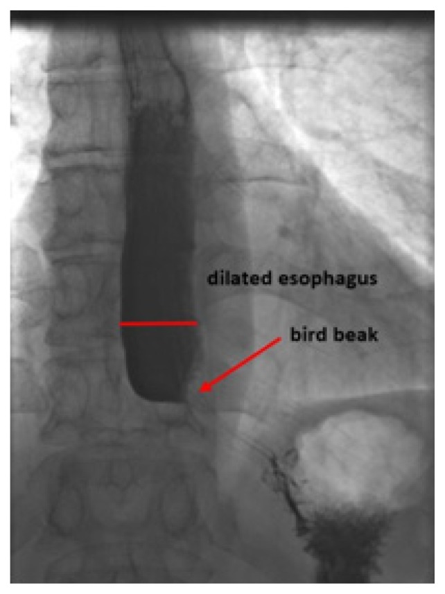
Figure 2.
Initial high resolution esophageal manometry image documenting segmental aperistalsis (A) in the distal esophagus from about 6cm above the LES, normal propagation of the peristaltic wave (B) with a normal distal latency (DL) of 5.85s (normative >4.5s) and a poorly relaxing lower esophageal sphincter (LES) defined by an integrated relaxation pressure (IRP4s) of 20.2 mmHg (normative <17 mmHg in achalasia type III) and a residual pressure of 21 mmHg (normative <8mmHg) with a slightly elevated resting pressure of 46mmHg (normative 10–45 mmHg).

