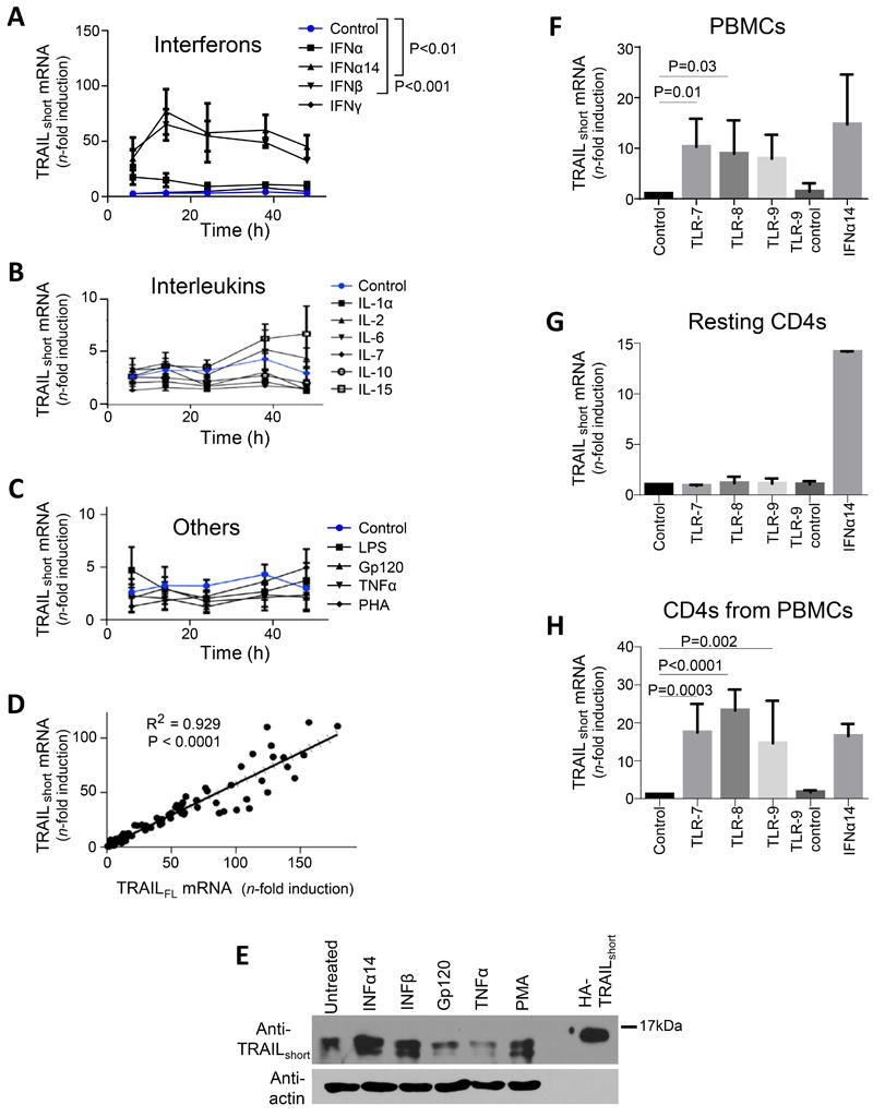
Figure 2. Type I interferons and TLR agonists drive production of TRAILshort.

(A, B, and C) Primary uninfected resting (CD25−, CD69−, HLA-DR−) CD4 T cells were either unstimulated, or stimulated with interferons (A), interleukins (B), or other biologically active proteins (C) and TRAILshort mRNA measured by qRT-PCR. (D) Concomitant TRAIL and TRAILshort mRNA expression was compared across samples. (E) Resting CD4 T cells were stimulated as depicted and TRAILshort protein expression assessed by western blot. Representative blot is shown from three independent experiments. (F) PBMCs were treated with vehicle control, TLR 7, 8, or 9 agonists or TL9 inactive control for 24 hours and TRAILshort mRNA expression measured. (G) Resting isolated CD4 T were treated similarly as the PBMCs in (F) and TRAILshort mRNA measured. (H) PBMCs were treated as in (F), and after 24 hours, CD4 T cells were separated and TRAILshort mRNA measured in the CD4 subset. Data represent means (SEM) of 5 independent experiments per treatment. P<0.05 considered statistically significant.
