Fig. 2.

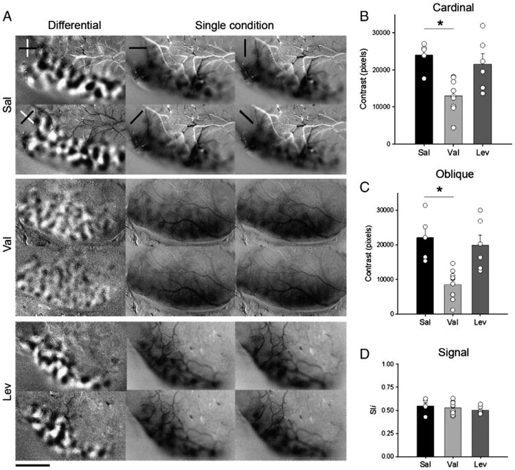
Early exposure to Val disrupts orientation selectivity. A. Analysis of orientation selectivity by optical imaging of intrinsic signals of juvenile (P42–P56) ferrets, submitted to different treatments (Sal, Val and Lev). In cardinal differential maps (first column and row from each experimental group), dark and light areas represent regions that respond preferentially to 0° (horizontal) and 90° (vertical) gratings respectively. In oblique differential maps (first column and second row from each experimental group), dark and light areas represent regions that respond preferentially to 45° and 135° gratings respectively. Note the poor contrast in both cardinal and oblique differential maps in the Val-treated animals relative to the Sal and Lev-treated animals. In single condition maps, dark areas represent regions responsive to a single angle. Stimulus orientations used were 0°, 45°, 90°, and 135°. Note that robust responses were present at every orientation tested. Scale bar = 3 mm. Quantification of contrast differences displayed for cardinal (B) and oblique (C) differential maps of Sal- Val- and Lev-treated groups. *P<0.01. Bars represent means (± SEM), and circles represent individual animal values. Note that most of the animals in the Val-group have a lower contrast than controls, while the animals in the Lev group present contrast values similar to Sal ones. D. Signal strength measured by pixel intensity. Note the similarity between groups.
