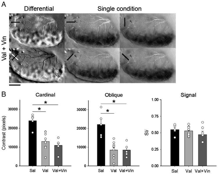
Fig. 5.

Vinpocetine does not restore orientation selectivity after early exposure to Val. A. Representative case of cardinal and oblique differential maps and single condition maps in Val+Vin-treated animals, as revealed by optical imaging of intrinsic signals. Note that, poor contrast maps can still be seen in Val + Vin treated animal after giving vinpocetine. Single condition maps show robust response at every orientation tested. Scale bar = 2.5 mm. Quantification of contrast differences (B) displayed for cardinal, oblique differential maps, and signal of Sal, Val and Val + Vin-treated groups. *P<0.01. Bars represent means (± SEM), and circles represent individual animal values. Note that animals in the Val and Val + Vin-group have a lower contrast than controls. Signal strength measured by pixel intensity. Note the similarity between groups.
