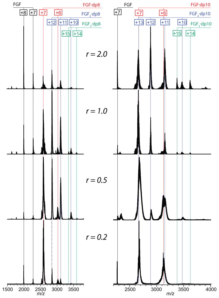
Figure 2.
ESI mass spectra of FGF-1 incubated with dp8 (left panel) and dp10 (right panel) it 100 mM NH4CH3CO2 at pH 6.8. The numbers in boxes indicate charge states of free FGF-1 (black), and its complexes with heparinoids: 1:1 (red), 2:1 (blue) and 3:1 (green). The r values shown in each row indicate the protein/heparinoid molar ratio.

