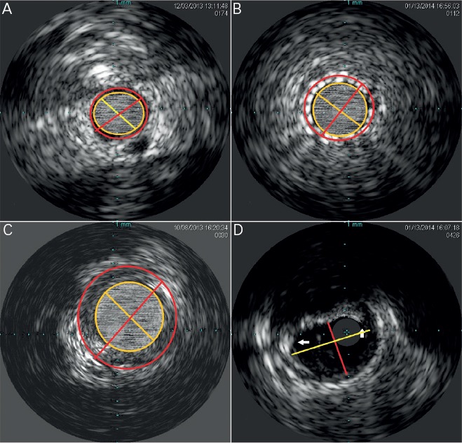
Figure 2: Basic Intravascular Ultrasound Measurements.

A) The lumen CSA is the area contained within the yellow inner circle. The EEM CSA is contained by the outer red circle. The yellow line represents the lumen diameter, and the red line represents the total vessel diameter. B) The stent CSA is the area contained within the yellow inner circle. The EEM CSA is contained by the outer red circle. The yellow line represents the stent lumen diameter, and the red line represents the total vessel diameter. C) After excluding the lumen CSA from the EEM CSA the residual area represents the atheroma CSA (Plaque and media CSA. D) The maximum luminal diameter is represented by the longer yellow line, and the minimum lumen diameter is represented by the shorter red line. The white arrows point to the borders of the near 155 degree arc of fibro-calcific plaque. CSA = cross sectional area; EEM = External elastic membrane.
