
Extended Data Figure 4. Distribution and outcome of 28 HER2 exon 20 insertions.
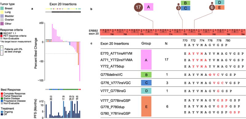
a) Percent best change and PFS plots corresponding to each type of exon 20 insertion (colour coded by synonymous amino acid change). Three cases with no change are indicated in colour-coded circles above the x-axis. b) Zoomed-in schematic of all exon 20 insertions positioned by their amino acid co-ordinates and frequencies. c) Five unique types of exon 20 insertions observed in the study with the resulting full amino acid sequences (insertion indicated in red).
PET, positron-emission tomography; PFS, progression-free survival; RECIST, Response Evaluation Criteria in Solid Tumors.