Figure 1.
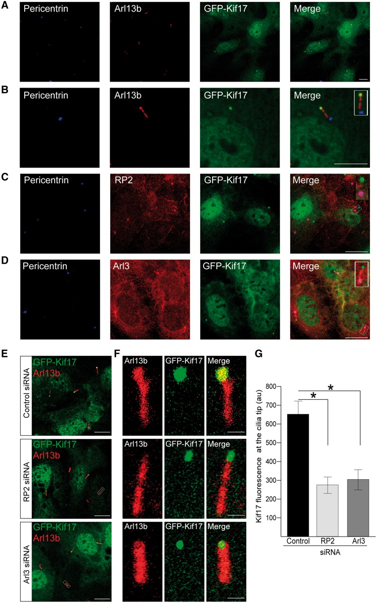
RP2 and Arl3 mediate GFP-Kif17 cilia tip localisation. (A) In stable GFP-Kif17 hTERT-RPE cells Kif17 (green) localises to the nucleus, the cytoplasm and to cilia tips, as indicated by the cilia marker Arl13b (red) and the basal body marker pericentrin (blue). Scale bar 10 μm. (B) Zoomed image of GFP-Kif17 hTERT-RPE cell line. Inset shows a zoomed in image of the cilia tip. Scale bar 10 μm. (C) RP2 (red) localises to the basal body (pericentrin, blue) of cilia in GFP-Kif17 hTERT-RPE cells. Scale bar 10 μm. (D) Arl3 (red) localises to the basal body (pericentrin, blue) and the along the ciliary axoneme in GFP-Kif17 hTERT-RPE cells. Scale bar 10 μm. (E) siRNA-mediated knockdown of RP2 or Arl3 decreases GFP-Kif17 at cilia tips. Cilia marker Arl13b (red). Scale bar 10 μm. (F) Zoomed image of GFP-Kif17 at cilia tips following control, RP2 or Arl3 siRNA transfection. Cilia marker Arl13b (red). Scale bar 1 μm. (G) Quantification of GFP-Kif17 fluorescence at cilia tips after control, RP2 or Arl3 siRNA transfection. n = 3 independent experiments. *P ≤ 0.05, values are mean ± SEM.

