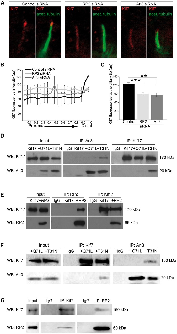
Figure 2.
RP2 and Arl3 mediate the localisation of Kif7 to cilia tips. (A) siRNA-mediated knockdown of RP2 or Arl3 in hTERT-RPE cells decreases Kif7 (red) at cilia tips. Cilia marker acetylated α-tubulin (green). Scale bar 1 μm. (B) Fluorescence intensity of Kif7 along the axoneme, normalised for cilium length. n = 3 independent experiments, with a total of 90 cilia measured for each siRNA treatment. For RP2 siRNA, P ≤ 0.0001 for the last measurement of the cilia tip. For Arl3 siRNA, P ≤ 0.05 and P ≤ 0.001, for the distal part of cilia. Values are mean ± SEM. (C) Quantification of Kif7 fluorescence at cilia tips. n = 3 independent experiments, with a total of 90 cilia measured for each siRNA treatment. ***P ≤ 0.0001, **P ≤ 0.001, values are mean ± SEM. (D-G) Reciprocal co-IPs of different combinations; (D) GFP-Kif17 with the myc-tagged Arl3 conformational mimics Arl3-GTP (Q71L) and Arl3-GDP (T31N) shows that GFP-Kif17 preferentially binds to Arl3-T31N. (E) GFP-Kif17 binds to RP2. (F) V5-Kif7 with the myc-tagged Arl3 conformational mimics Arl3-GTP (Q71L) and Arl3-GDP (T31N) shows that Kif7 preferentially binds to Arl3-T31N. Kif7 and Arl3 were immunopurified with the V5 and myc antibody, respectively. (G) V5-Kif7 binds to RP2. Kif7 and RP2 were immunopurified with the V5 antibody and GFP-trap magnetic beads, respectively.

