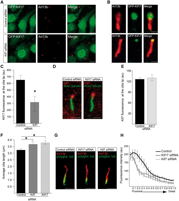
Figure 3.
Kif7 functions upstream of Kif17 in cilia and both kinesins contribute to cilia stability. (A) siRNA-mediated knockdown of Kif7 in GFP-Kif17 hTERT-RPE cells reduces GFP-Kif17 at cilia tips. Scale bar 10 μm. (B) Zoomed in image of GFP-Kif17 cilia localisation following control or Kif7 siRNA treatment. Scale bar 1 μm. (C) Quantification of GFP-Kif17 fluorescence at the cilia tip following siRNA treatment. n = 3 independent experiments with 150 cilia measured for each siRNA. *P ≤ 0.05, values are mean ± SEM. (D) hTERT-RPE cells stained for Kif7 (red) and acetylated α-tubulin (green) following siRNA treatment. Scale bar 1 μm. (E) Quantification of Kif7 fluorescence at the cilia tip following siRNA treatment. n = 3 independent experiments with 150 cilia analysed for each siRNA. (F) Cilia length in hTERT-RPE cells treated with Kif7 and Kif17 siRNA is increased compared to controls. n = 3 independent experiments with 150 cilia measured for each siRNA. *P ≤ 0.05, values are mean ± SEM. (G) hTERT-RPE cilia treated with Kif17 or Kif7 siRNA show decreased levels of polyglutamylated tubulin (green) compared to controls. Cilia marker, Arl13b (red). Scale bar 1 μm. (H) Fluorescence intensity of polyglutamylated tubulin, normalised for cilium length. n = 3 independent experiments, with a total of 90 cilia measured for each siRNA treatment. For Kif7 and Kif17 siRNA, P ≤ 0.05 for the proximal part of cilia. Values are mean ± SEM.

