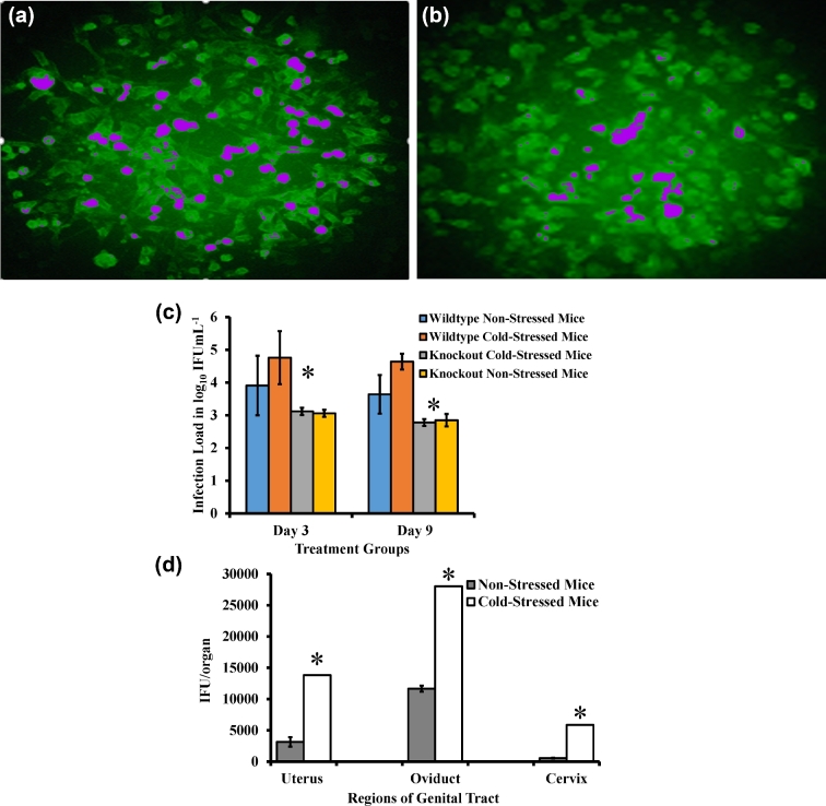
Figure 3.
Determination of C. muridarum shedding in the genital tract of wild type and knockout stressed mice. Stressed (a) and non-stressed (b) mice were infected with C. muridarum. Cervicovaginal swabs were collected at 3-day intervals (c). Alternately, cervix, uterus and oviduct sections were harvested after 48 h of infection (d). McCoy mouse fibroblasts were seeded into 96-well plates and incubated at 37°C with 5% carbon dioxide supply until monolayers were 90%–95% confluent. After 32 h of infection, C. muridarum isolation was performed by staining with fluorescence-tagged anti-chlamydia antibodies and inclusion bodies were counted. Each data point is mean ± standard deviation of log10 inclusion forming unit/milliliter representing combined results of two separate experiments (n = 5 to 6 mice per group). False color has been added to (a) and (b) to aid visualization of the inclusion bodies.

