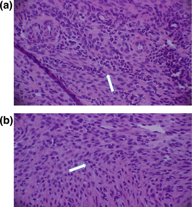
Figure 4.

Infiltration of leukocytes to the regions of genital tract of mice during C. muridarum genital infection. Arrows show neutrophils the cervix of stressed mice (a) and non-stressed mice (b) during C. muridarum infection. Mice were stressed, infected as above. After 48 h of infection, mice were euthanized and the tract was aseptically harvested and excised into cervix, uterus and oviduct sections and placed in buffered formalin. The sections were stained with hematoxylin and eosin (H&E) at Marshall University pathology laboratory services.
