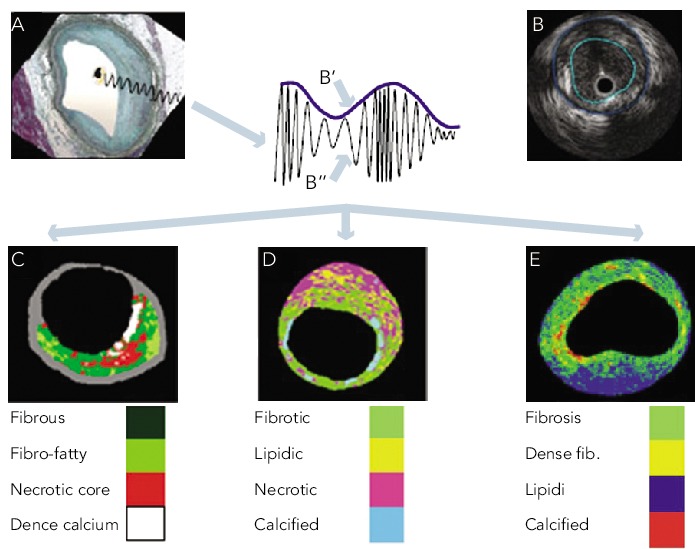
Figure 1: Grey-scale Intravascular Ultrasound and Intravascular Ultrasound Radiofrequency Analysis.

A. An IVUS is obtained from the vessel wall within an histology image. B. The greyscale IVUS image is formed by the envelope (amplitude) of the radiofrequency signal. C. From the backscatter radiofrequency data different types of tissue information can be retrieved: virtual histology; iMAP (D) and integrated backscattered IVUS (E). Adapted from Garcìa-Garcìa HM et al.[4]
